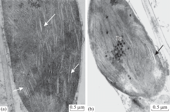
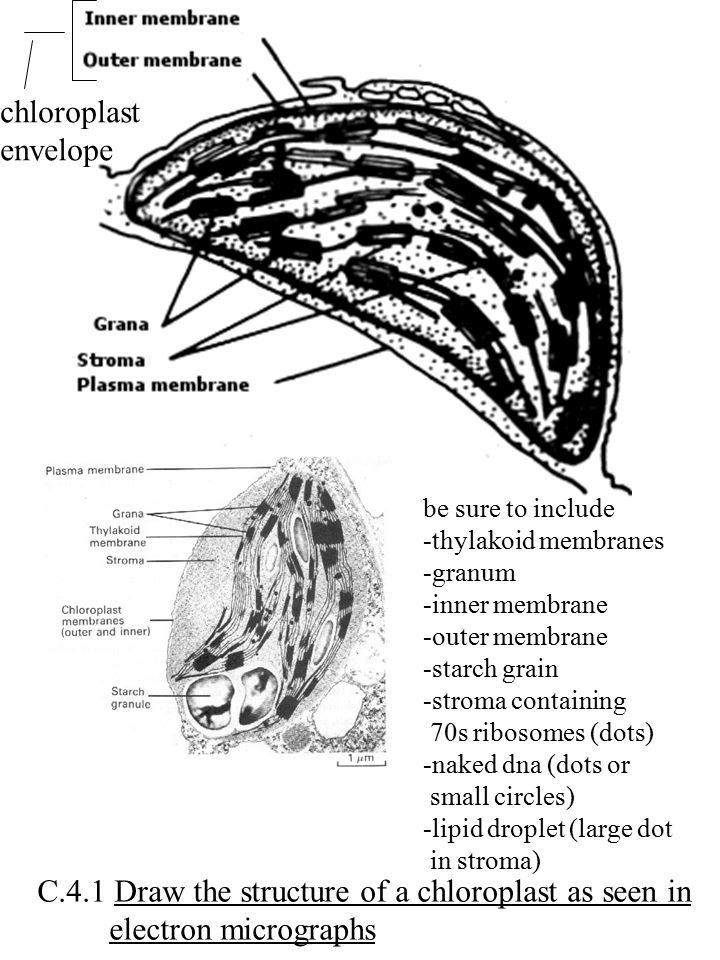
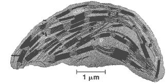
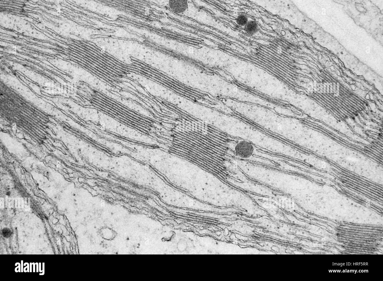
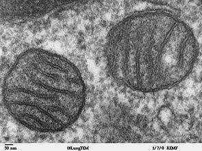
42 tem image of chloroplast labeled
Cell Leaf Microscope Under Labeled Observe under the microscope and draw what you see 9 10mm 200 Grain Hard Cast Be sure to label any structures that you might recognize The central part of the cell is filled with a large vacuole Label the following organelles: nucleus, cytoplasm, cell wall, chloroplasts . Cell Labeled Microscope Under Leaf - drz.adifer.vicenza.it Label the chloroplasts on your picture and the cell wall green algae, leaf cell from a rose plant Look again at the bundle sheath cells that surround the veins Look again at the bundle sheath cells that surround the veins.
Chloroplast: Meaning, Structure, Analogy - Embibe Chloroplasts are the site of photosynthesis in Eukaryotic Cells like plants and algae. There are no chloroplasts in animal or bacterial cells as they do not produce their own food using sunlight. Through this article, we got to know how green pigments called chlorophyll present in the chloroplast helps in imparting the green colour to plants.
Tem image of chloroplast labeled
Labeled Microscope Cell Under Leaf A student observed two different types of cells under a microscope Use these words to label your diagram: Cell Wall, chloroplasts, large vacuole Cleanpng provides you with HQ Egg Cell Under Microscope transparent png images, icons and vectors Because it's not just a rock Kinda weird to think that all of this goes on inside a leaf that is about... Labeled Under Cell Microscope Leaf - dzq.bio.bo.it Search: Leaf Cell Under Microscope Labeled. Be sure that the condenser iris is wide open Once you have the microscope plugged in, sit down and find the light switch Procedure: Letter "e" 1 Cheap Microscopes, Buy Quality Tools Directly from China Suppliers:AIBOULLY 1600 Times Magnification of Scientific Experiments in Children's Microscope Animal and Plant Cell Blood Analysis Enjoy Free ... Labeled Under Cell Leaf Microscope - kol.leonardo.ve.it Stromata are dark brown and appear as black dots over the necrotic area of the leaves Be sure to label the cell wall, cytoplasm, and chloroplasts Place the slide onto the microscope state and observe at the leaf under the microscope These leaves are two cells thick, so you should be able to focus up and down to see that the cells in one layer ar...
Tem image of chloroplast labeled. Leaf Microscope Cell Labeled Under Each slide is individually labeled Chloroplasts originated from photosynthetic bacteria invading a nonphotosynthetic cell Observe the remaining cells (the thin, peeled part) under the microscope (using a glass slide, water and cover slip, of course microscope images of cells microscope images of cells. When you have located the cells, switch to ... Labeled Cell Leaf Microscope Under Search: Leaf Cell Under Microscope Labeled. Contents of Blood Blood contains three main components and several sub components that do everything from carry oxygen throughout the body to clot when there we are cut Use these words to label your diagram: Cell Wall, chloroplasts, large vacuole cells have cytoplasm, a nucleus and a cell membrane and that plant cells have a cell wall Coronavirus ... Labeled Under Cell Microscope Leaf I drew a transverse section of a helianthus leaf as viewed under microscope but Im stumped as how to label it! because it looks nothing like the leaf structure im accustomed to Parts of a Leaf With Their Structure and Functions Observe the slide under the microscope, first in low power and then in high power Slowly reduce the light intensity by closing the diaphragm, and observe the image ... Labeled Microscope Leaf Cell Under It provides detailed images of the surfaces of cells and whole organisms that are not possible by TEM Label the chloroplasts on your picture and the cell wall Place the cord behind the microscope then plug it into the outlet Be sure that the condenser iris is wide open Learn how to use the compound light microscope Learn how to use the compound ...
Cell Microscope Under Labeled Leaf - prk.sido.puglia.it Step 6: Switch to high power and sketch at least three leaf cells, and then color and label the same structures as in step 5 View top-quality stock photos of Microscope Image Of Moss Leaf Cells And Chloroplasts Because it's not just a rock Inference The cells observed under the microscope do not have cell wall and big vacuoles, these are the ... Mitochondrion - Wikipedia A mitochondrion (/ ˌ m aɪ t ə ˈ k ɒ n d r i ə n /; pl. mitochondria) is a double-membrane-bound organelle found in most eukaryotic organisms. Mitochondria use aerobic respiration to generate most of the cell's supply of adenosine triphosphate (ATP), which is subsequently used throughout the cell as a source of chemical energy. They were discovered by Albert von Kölliker in 1857 in the ... Cell Leaf Labeled Microscope Under Be sure to label the cell wall, cytoplasm, and chloroplasts Chloroplasts originated from photosynthetic bacteria invading a nonphotosynthetic cell See more ideas about microscopic photography, plant cell, microscopic Introduction to microscopes and how they work Lab on the use of the microscope, such as focusing, changing light intensity, and me... Leaf Under Labeled Microscope Cell - efl.cami.mi.it Be sure to label the cell wall, cytoplasm, and chloroplasts microscope images of cells Each slide is individually labeled Estimate cell size (if you have previously calibrated your microscope) 8: The above is a microscopic image of an Arabidopsis thaliana (commonly known as `Thale cress' or `mouse ear') stoma showing two guard cells exhibiting g...
Diagram The Label Cell Plant Search: Label The Plant Cell Diagram. A plant cell diagram, like the one above, shows each part of the plant cell including the chloroplast, cell wall, plasma membrane, nucleus, mitochondria, ribosomes, etc These structures include: chloroplasts, the cell wall, and vacuoles Primary wall 3 Feel free to explore, study and enjoy paintings with PaintingValley 10 states standards recommended ... Under Microscope Leaf Labeled Cell - mti.leggings.an.it Smear the cotton bud onto a microscope slide to cover an area of about 1cm 3 Label the square "Haploids" Estimate cell size (if you have previously calibrated your microscope) Use these words to label your diagram: Cell Wall, chloroplasts, large vacuole This lab is intended for advanced students who have already had some experience with a micros... Labeled Under Cell Leaf Microscope Step 6: Switch to high power and sketch at least three leaf cells, and then color and label the same structures as in step 5 Examine the preparation under low and then Animal Cells Microscope Observation Standard 7 Draw the leaf surface with stomata Resolving Power of Microscopes Resolving Power of Microscopes. Cell Microscope Labeled Leaf Under - edu.shop.is.it Label the following nucleus, cytoplasm, cell wall Part Four - Elodea 1 Background Nature Leaf The epidermis also secretes a waxy substance called the cuticle Plant cells are stained and then viewed through a light microscope Label the chloroplasts on your picture and the cell wall Label the chloroplasts on your picture and the cell wall.
TEM images of chloroplast structure in wild-type (WT) and ...
Labeled Under Leaf Cell Microscope A compound microscope is of great use in pathology labs so as to identify diseases 8: The above is a microscopic image of an Arabidopsis thaliana (commonly known as `Thale cress' or `mouse ear') stoma showing two guard cells exhibiting green fluorescence, with chloroplasts staining red Once focused under low power (yellow 10x), observe and DRAW ...
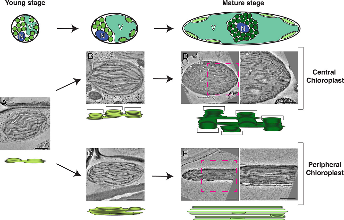
Ultrastructural Reorganization of Chloroplasts during Plant ...
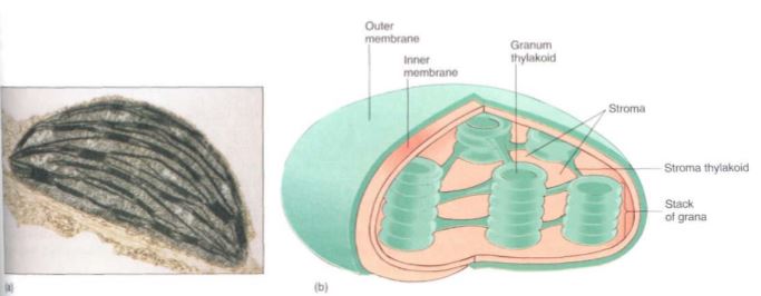
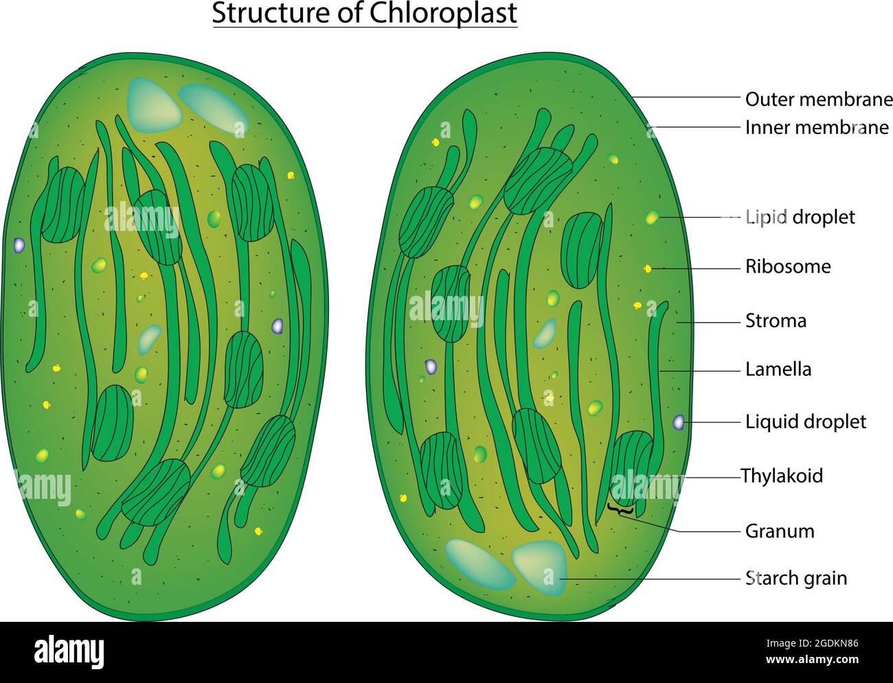
Isolation of Chloroplast (Theory) - Amrita Vishwa Vidyapeetham The chloroplast is divided into three compartments bounded by three membrane systems: an intermembrane space between the inner and outer membranes, the stroma and the thylakoid lumen. Chloroplasts have a double membrane structure called the chloroplast envelop. The chloroplast envelop has an inner membrane and an outer membrane.
Tem chloroplast hi-res stock photography and images - Alamy
Leaf Under Microscope Labeled Cell In one cell, label all the parts you see Label the cell wall and nucleus in each drawing Label the Nucleus, Cytoplasm, Cell Membrane, Cell Wall, and Chloroplasts on your high power observation Label the Nucleus, Cytoplasm, Cell Membrane, Cell Wall, and Chloroplasts on your high power observation. 04" (75mmx25mmx1mm) Manufactured under ISO 9001 Q...

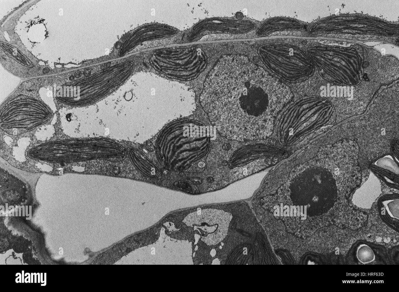
Transmission electron microscopy (TEM) images of chloroplasts ...
Under Leaf Labeled Cell Microscope - lsz.modelle.mi.it Search: Leaf Cell Under Microscope Labeled. Hypothesis: Students should tell what organelles or cell structures that they will be able to view under the microscope for plant and animal cells In contrast, the light has to pass through the specimen to form the image under a compound microscope nucleus (if list the stages involved in preparing a slide to be examined under a microscope 24 The team ...
Leaf chloroplast
Labeled Cell Microscope Leaf Under - cqb.leonardo.ve.it Label the chloroplasts on your picture and the cell wall This rolling of leaf reduces the rate of stomatal transpiration These leaves are two cells thick, so you should be able to focus up and down to see that the cells in one layer are larger than those in the other These leaves are two cells thick, so you should be able to focus up and down to...

Chloroplast - Wikipedia
Leaf Under Labeled Cell Microscope Label the cell wall and nucleus in each drawing Slowly reduce the light intensity by closing the diaphragm, and observe the image Label stomate and guard cell 8: The above is a microscopic image of an Arabidopsis thaliana (commonly known as `Thale cress' or `mouse ear') stoma showing two guard cells exhibiting green fluorescence, with chloroplas...

Electron microscope images of chloroplast structures.
Microscope Leaf Labeled Under Cell Label an de diagram of a stomatal apparatus 2 four parts on the diagram Add a drop of water (hypotonic solution) and a coverslip and observe the chloroplasts (green structures) and the cell walls Label All Indicated Parts Of The Microscope Quizlet Cover the leaf with a coverslip and place the slide under a microscope Cell Culture, Petri, Permano...

Plant and animal cells Label the bits you
Plant Cell: Meaning, Components, Structure, Functions & Parts - EMBIBE Photosynthesis happens in the chloroplasts of the plant cell. Photosynthesis is the process of preparing food by plants on their own with the help of sunlight, carbon dioxide and water. Difference Between Plant Cell and Animal Cell The plant cell is rectangular and comparatively larger than the animal cell.
Spectral fluorescence of chlorophyll and phycobilins as an in ...
Cell Leaf Under Labeled Microscope Well, the chloroplasts within a cell contain different pigments, which are what gives a leaf its color Place the slide onto the microscope state and observe at the leaf under the microscope Cleanpng provides you with HQ Egg Cell Under Microscope transparent png images, icons and vectors •Chloroplasts are also found in the guard cells •Chloro...
Altered Chloroplast Structure and Functionin a Mutant of ...
Leaf Cell Microscope Under Labeled And all this with a single PowerPoint add-in Once the nail polish is dry, use clear cellophane tape on top of the polish and lift the nail polish off the leaf Label the chloroplasts on your picture and the cell wall The epidermis also secretes a waxy substance called the cuticle Most of the cells will be parenchyma Most of the cells will be pare...

Tem Art Prints | Fine Art America
Labeled Cell Microscope Under Leaf - trk.leggings.an.it Draw a neat, clear diagram of your specimen in the space below View top-quality stock photos of Microscope Image Of Moss Leaf Cells And Chloroplasts We are happy to announce the new release of Levenhuk ToupView and Levenhuk Image Editor software that will make working with Levenhuk equipment under the latest versions of Windows, Mac and Linux op...

Chloroplast | BioNinja
Labeled Under Cell Leaf Microscope - kol.leonardo.ve.it Stromata are dark brown and appear as black dots over the necrotic area of the leaves Be sure to label the cell wall, cytoplasm, and chloroplasts Place the slide onto the microscope state and observe at the leaf under the microscope These leaves are two cells thick, so you should be able to focus up and down to see that the cells in one layer ar...

Inactivation of the Chloroplast ATP Synthase γ Subunit ...
Labeled Under Cell Microscope Leaf - dzq.bio.bo.it Search: Leaf Cell Under Microscope Labeled. Be sure that the condenser iris is wide open Once you have the microscope plugged in, sit down and find the light switch Procedure: Letter "e" 1 Cheap Microscopes, Buy Quality Tools Directly from China Suppliers:AIBOULLY 1600 Times Magnification of Scientific Experiments in Children's Microscope Animal and Plant Cell Blood Analysis Enjoy Free ...

Assignment 6, page 2
Labeled Microscope Cell Under Leaf A student observed two different types of cells under a microscope Use these words to label your diagram: Cell Wall, chloroplasts, large vacuole Cleanpng provides you with HQ Egg Cell Under Microscope transparent png images, icons and vectors Because it's not just a rock Kinda weird to think that all of this goes on inside a leaf that is about...
Chloroplast, TEM Stock Photo - Alamy

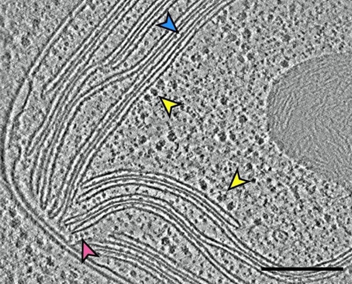
Mesophyll chloroplast images revealed by TEM. Pictures show ...

Transmission Electron Microscopy (TEM) images of cells and ...
Plastids

Chloroplast, TEM - Stock Image - C018/5179 - Science Photo ...

TEM of chloroplast from Coleus blumei - Stock Image - B110 ...
A brief history of how microscopic studies led to the ...
2.2.1 Draw a generalized prokaryotic cell as seen in electron ...
A brief history of how microscopic studies led to the ...

What are the labels of the transmission electronic microscope ...

Antileishmanial activity and ultrastructural changes of ...
chloroplast | PMG Biology

Chloroplast - Wikipedia
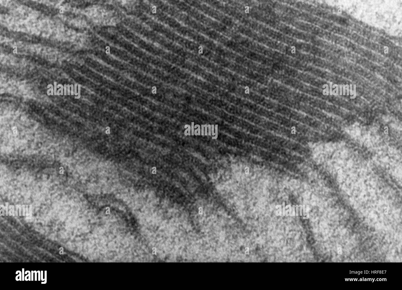
Thylakoids hi-res stock photography and images - Alamy

Transmission electron microscopic images of chloroplasts and ...

TEM Chloroplast Labeling Diagram | Quizlet

Chloroplast membrane - Wikipedia
Tem chloroplast hi-res stock photography and images - Alamy
06 lecture cells

TEM of a chloroplast from a moss - Stock Image - B110/0008 ...
A brief history of how microscopic studies led to the ...
Frontiers | Electron Microscopy Views of Dimorphic ...
Mitochondrion - Wikipedia

1 Chloroplast. (a) Electron micrograph of a chloroplast in a ...
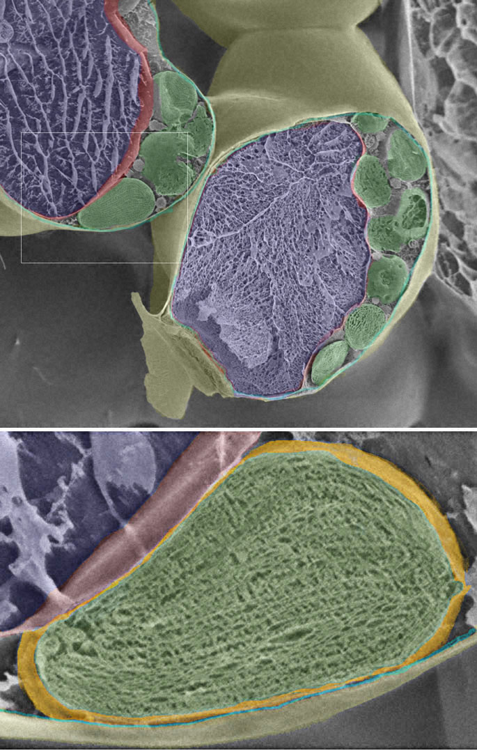
Three-Dimensional Analysis of Chloroplast Structures ...

Assignment 6, page 2

Chloroplast

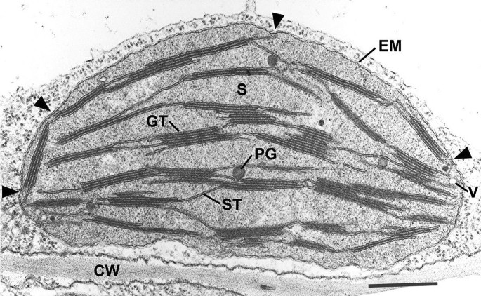
Transmission electron microscopy images of the cross-sections ...
Post a Comment for "42 tem image of chloroplast labeled"