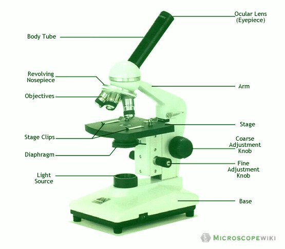
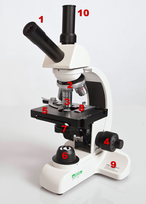
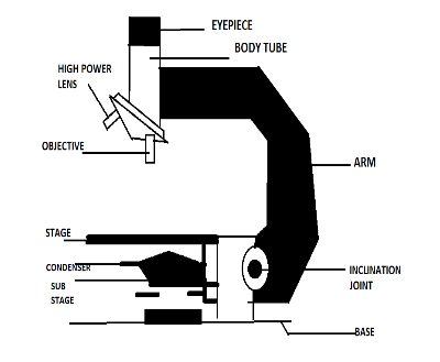
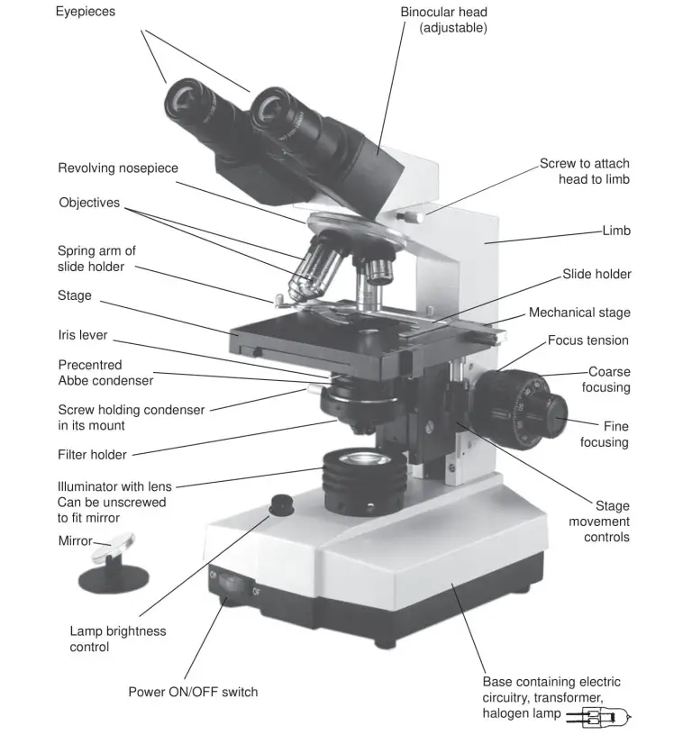
43 a well labelled diagram of a binocular microscope
dictionary.cambridge.org › us › dictionaryWELL | definition in the Cambridge English Dictionary As well (as) meaning ‘in addition’. As well is an adverb which means ‘also’, ‘too’ or ‘in addition’. We usually use as well at the end of a clause: …. Might as well and may as well. We use might as well and may as well informally to mean that something is worth doing only because other things are not happening. What Are The Different Parts of a Binocular? - The Ultimate Guide!! Yes, the answer is the Binoculars two-barrel chambers that fit these parts together and in alignment. Both the barrels of a binocular need to be optically parallel for the image to merge into one perfect circle. Also. the two barrels remain aligned with each other no matter what the gap between the pupils of your eyes.
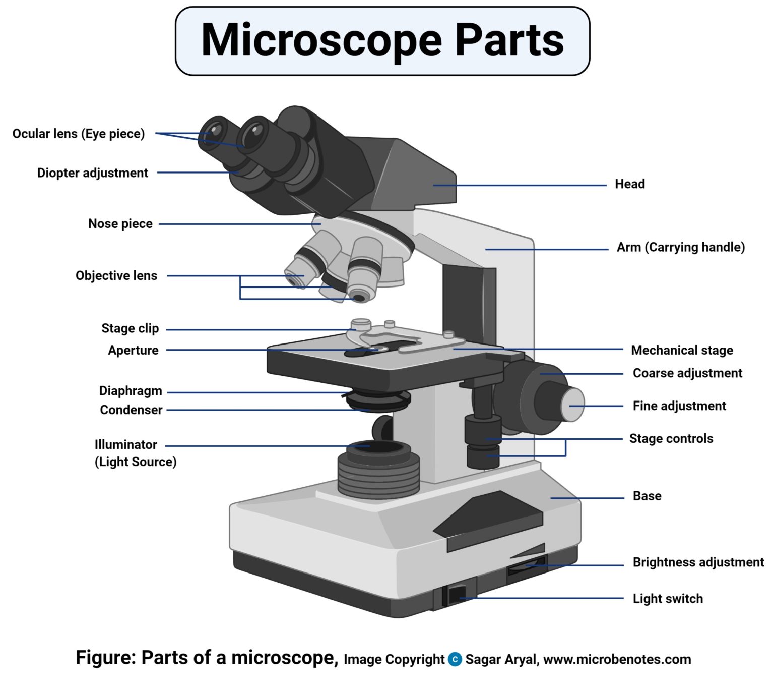
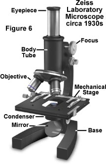
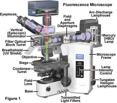
Parts of a microscope with functions and labeled diagram - Microbe Notes Parts of a microscope with functions and labeled diagram September 17, 2022 by Faith Mokobi Having been constructed in the 16th Century, Microscopes have revolutionalized science with their ability to magnify small objects such as microbial cells, producing images with definitive structures that are identifiable and characterizable.
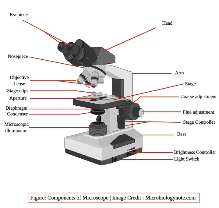
A well labelled diagram of a binocular microscope
Diagram of a Compound Microscope - Biology Discussion Diagram of a Compound Microscope Article Shared by ADVERTISEMENTS: In this article we will discuss about:- 1. Essential Parts of Compound Microscope 2. Magnification of the Image of the Object by Compound Microscope 3. Resolution Power 4. Method for Studying Microbes 5. Measurement of the Size of Objects. Essential Parts of Compound Microscope: PDF Label parts of the Microscope: Answers Label parts of the Microscope: Answers Coarse Focus Fine Focus Eyepiece Arm Rack Stop Stage Clip . Created Date: 20150715115425Z ... › piedmontPiedmont Urgent Care by WellStreet | Urgent Care in Georgia 2701 Holcomb Bridge Rd. 770-998-7611. Mon - Fri: 8am - 8pm. Sat - Sun: 8am - 8pm. Location Info. Book Ahead. Virtual Visit. Sandy Springs. 6660 Roswell Rd NE.
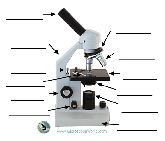
A well labelled diagram of a binocular microscope. Compound Microscope Parts, Functions, and Labeled Diagram ACCU-SCOPE 3000-LED Binocular Biological Microscope, 1000x Magnification $1,101.00 Meiji MT-30 Binocular Microscope - Rechargeable $618.55 Labomed 9135010 CxL Binocular Cordless Microscope, 4x, 10x, 40x Objectives, LED Illumination $713.21 ACCU-SCOPE EXM-150-MS Monocular Cordless Microscope with Mechanical Stage, Rechargeable $325.00 Labeling the Parts of the Microscope | Microscope World Resources Labeling the Parts of the Microscope. This activity has been designed for use in homes and schools. Each microscope layout (both blank and the version with answers) are available as PDF downloads. You can view a more in-depth review of each part of the microscope here. PaRTS of a BINOCULAR MICROSCOPE Flashcards | Quizlet one hand should grasp this part when carrying the microscope coarse adjustment knob this part is used to move the body up and down closer to the specimen fine adjustment used for fine, detailed focusing of the microscope light control controls the intensity of the light base one hand should be under this part when microscope is carried What Are the Parts and Functions of a Binocular Microscope? The parts of a binocular microscope are the eye piece (ocular), mechanical stage, nose piece, objective lenses, condenser, lamp, microscope tube and prisms. Each part plays an important role in the microscope's function.
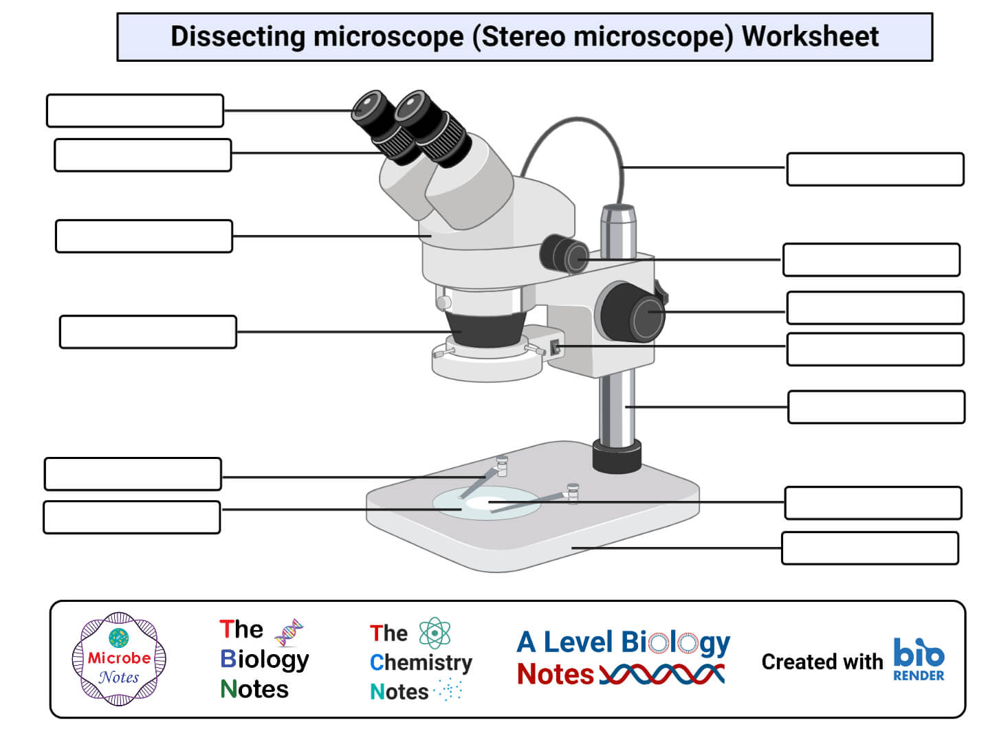
› es › translationwell - English-Spanish Dictionary - WordReference.com well adv (properly) bien adv : adecuadamente adv : The job has been done well. El trabajo se ha realizado bien. El trabajo se ha realizado adecuadamente. well adv (satisfactorily) bien adv : Things are going well lately; we have no unmet needs. The meeting went well, with no major difficulties. Las cosas están yendo bien últimamente; no nos está faltando nada. PDF Stereo Microscope - Home Science Tools Microscope Diagram Description of Components 1. Eye shields: These rubber shields fit over the eyepieces to block light and provide comfort. 2. Binocular eyepieces: This is the part of the microscope that you look through. The binocular eyepieces contain lenses that magnify 10x and provide an unreversed 3D stereo image. They are inclined at an ... › thesaurus › well507 Synonyms & Antonyms of WELL - Merriam-Webster noun. 1. as in source. a point or place at which something is invented or provided his quirkily dysfunctional family proved to be a bottomless well of inspiration for the novelist. Synonyms & Similar Words. source. cradle. Labelled Diagram Of A Light Microscope | Products & Suppliers ... The Radiant Vision Systems Microscope Lens enables high-resolution imaging of extremely small components and features, such as individual LEDs, display pixels, and subpixels. The lens attaches directly to a ProMetric (R) Imaging Colorimeter or Photometer, available in a range of resolutions.
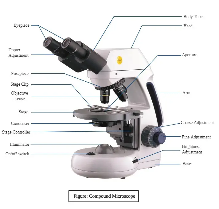
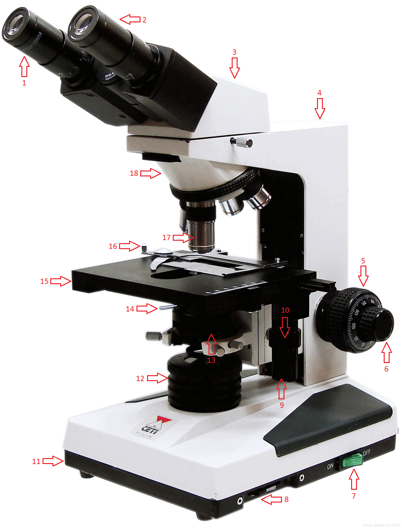
DOC Setting up a Binocular Microscope for Comfortable Viewing The goal of this part of the setup is to adjust the microscope so that your ciliary muscles are relaxed so that you can view microscopic specimens for hours without eye strain.) Using only your left eye, bring some small sharply defined portion of the specimen into sharp focus using the course and fine focus controls. Look up from the microscope. Compound Microscope - Diagram (Parts labelled), Principle and Uses The three structural components include: 1. Head - This is the upper part of the microscope that houses the optical parts. 2. Arm - This part connects the head with the base and provides stability to the microscope. Arm is used to carry the microscope around. 3. - International WELL Building Institute | IWBI WELL is an evidence-based roadmap for applying the WELL Building Standard to support the health and well-being of your people and your organization. Binocular Microscope Anatomy - Parts and Functions with a Labeled Diagram Now, I will discuss the details anatomy of the light compound microscope with the labeled diagram. Why it is called binocular: because it has two ocular lenses or an eyepiece on the head that attaches to the objective lens, this ocular lens magnifies the image produced by the objective lens. Binocular microscope parts and functions
Microscope Parts and Functions Microscope Parts and Functions With Labeled Diagram and Functions How does a Compound Microscope Work?. Before exploring microscope parts and functions, you should probably understand that the compound light microscope is more complicated than just a microscope with more than one lens.. First, the purpose of a microscope is to magnify a small object or to magnify the fine details of a larger ...
› wellWell - definition of well by The Free Dictionary well 2. (wɛl) n. 1. a hole drilled or bored into the earth to obtain water, petroleum, natural gas, brine, or sulfur. 2. a spring or natural source of water. 3. an apparent reservoir or a source of human feelings, emotions, energy, etc.: a well of compassion. 4. a container, receptacle, or reservoir for a liquid, as ink.
Microscope Parts, Function, & Labeled Diagram - slidingmotion Diaphragm. The diaphragm is also called as iris. This iris situates below the stage of the microscope. The function of the diaphragm is to control the amount of light that focuses on the specimen. This diaphragm can adjust the amount of light and intensity of light that falls on the specimen. In some standard and high-quality microscopes, this ...
Microscope Labeling Diagram | Quizlet Coarse Focus Knob Moves the stage large distances to roughly focus the image. Fine Focus Knob Moves the stage tiny distances to slightly adjust and fine-tune the image focus. Arm Supports the body tube. Objective Lenses Focus and magnify light in differing amounts to view the specimen. Stage Clips Hold the slide in place on the stage. Nosepiece
› Georgia › Find-a-ProviderFind a Provider | Wellcare Oct 1, 2021 · Find a Provider/Pharmacy. Who are you? Select type. . Select your state. Select your state. . Select your plan. Select your plan.
Compound Microscope Parts - Labeled Diagram and their Functions Labeled diagram of a compound microscope Major structural parts of a compound microscope Optical components of a compound microscope Eyepiece Eyepiece tube Objective lenses Nosepiece Specimen stage Coarse and fine focus knobs Rack stop Illuminator Condenser Abbe condenser Iris Diaphragm Condenser Focus Knob Summary An overview of microscopes
Compound Microscope- Definition, Labeled Diagram, Principle, Parts, Uses The optical microscope often referred to as the light microscope, is a type of microscope that uses visible light and a system of lenses to magnify images of small subjects. There are two basic types of optical microscopes: Simple microscopes. Compound microscopes. The term "compound" in compound microscopes refers to the microscope having ...
Labelled Diagram of Compound Microscope The below mentioned article provides a labelled diagram of compound microscope. Part # 1. The Stand: The stand is made up of a heavy foot which carries a curved inclinable limb or arm bearing the body tube. The foot is generally horse shoe-shaped structure (Fig. 2) which rests on table top or any other surface on which the microscope in kept.
2.4 Parts of the Petrographic Microscope - Introduction to Petrology There are often multiple apertures on a microscope (Figure 2.4.5), including an aperture as part of the condenser assembly, as well as a field diaphragm that controls the size of the area which is illuminated on a sample. Figure 2.4.7 shows the view through the microscope as the substage field diaphragm is opened and closed.
› piedmontPiedmont Urgent Care by WellStreet | Urgent Care in Georgia 2701 Holcomb Bridge Rd. 770-998-7611. Mon - Fri: 8am - 8pm. Sat - Sun: 8am - 8pm. Location Info. Book Ahead. Virtual Visit. Sandy Springs. 6660 Roswell Rd NE.
PDF Label parts of the Microscope: Answers Label parts of the Microscope: Answers Coarse Focus Fine Focus Eyepiece Arm Rack Stop Stage Clip . Created Date: 20150715115425Z ...
Diagram of a Compound Microscope - Biology Discussion Diagram of a Compound Microscope Article Shared by ADVERTISEMENTS: In this article we will discuss about:- 1. Essential Parts of Compound Microscope 2. Magnification of the Image of the Object by Compound Microscope 3. Resolution Power 4. Method for Studying Microbes 5. Measurement of the Size of Objects. Essential Parts of Compound Microscope:
















Post a Comment for "43 a well labelled diagram of a binocular microscope"