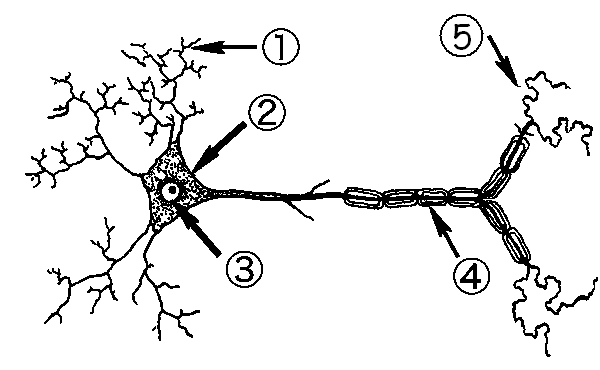
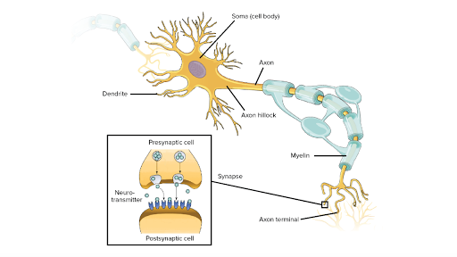
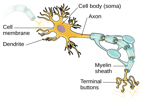
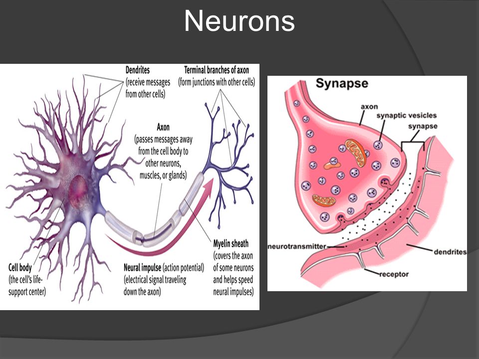
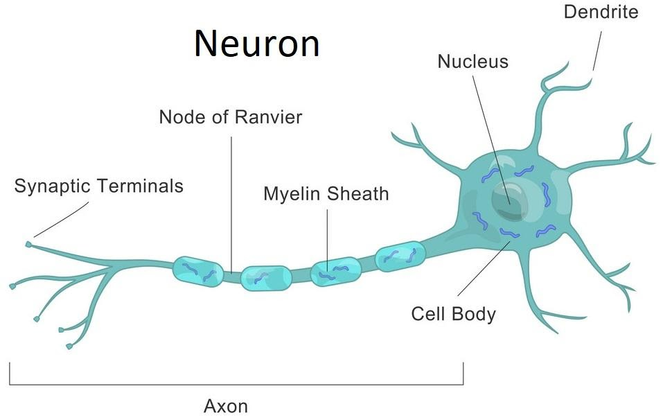
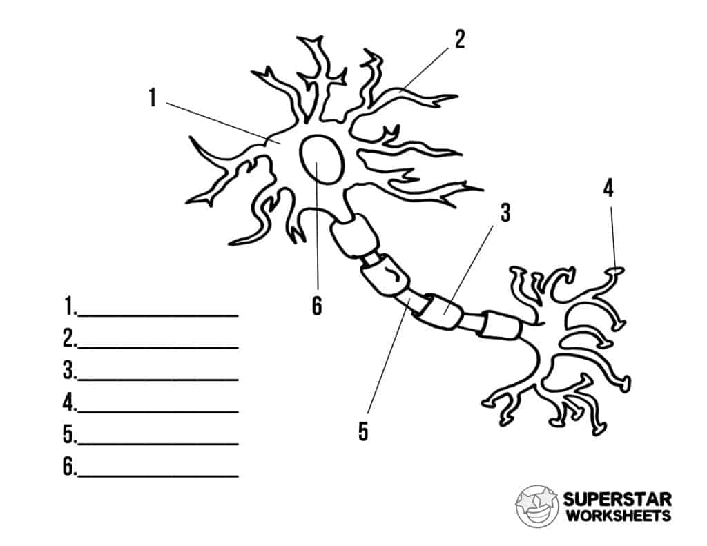
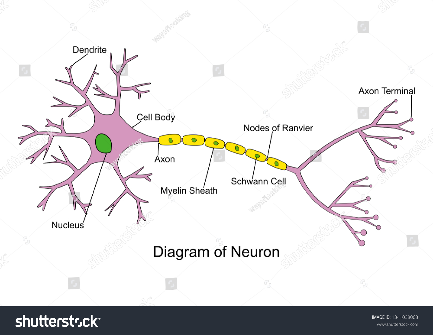
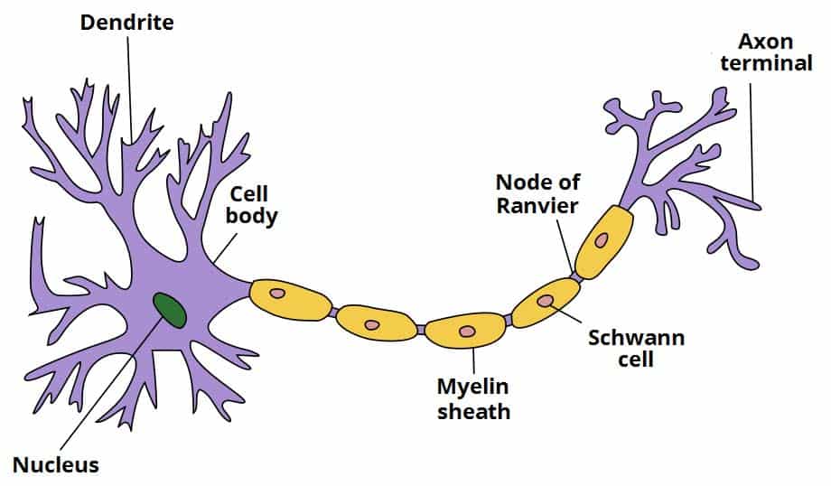
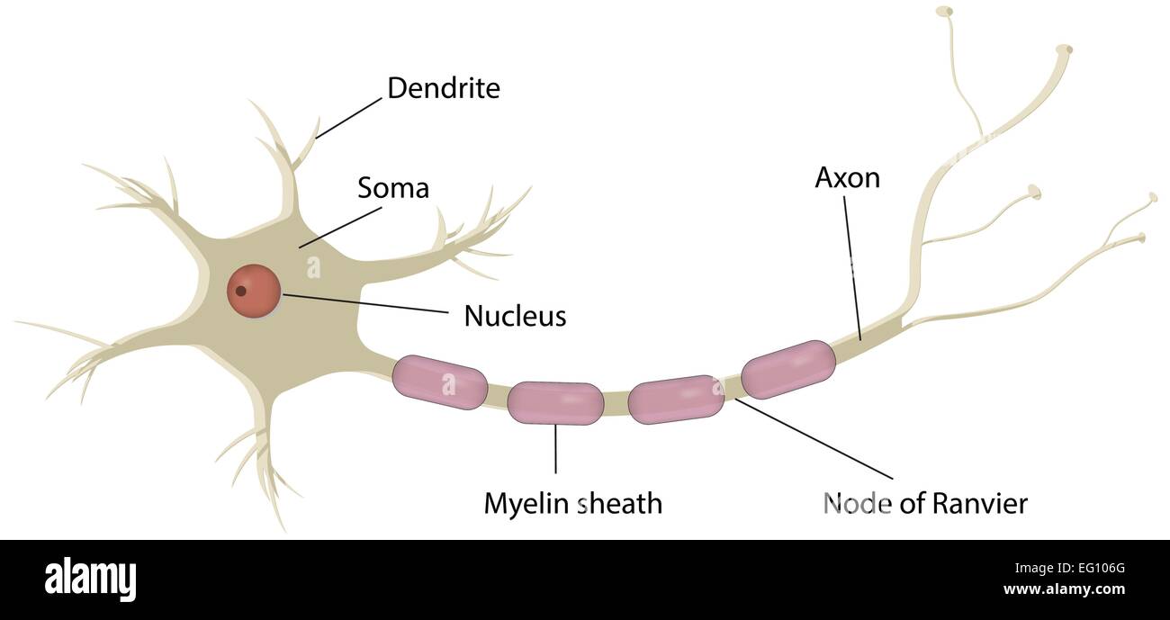
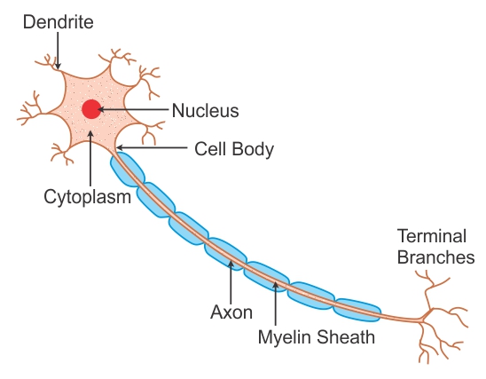
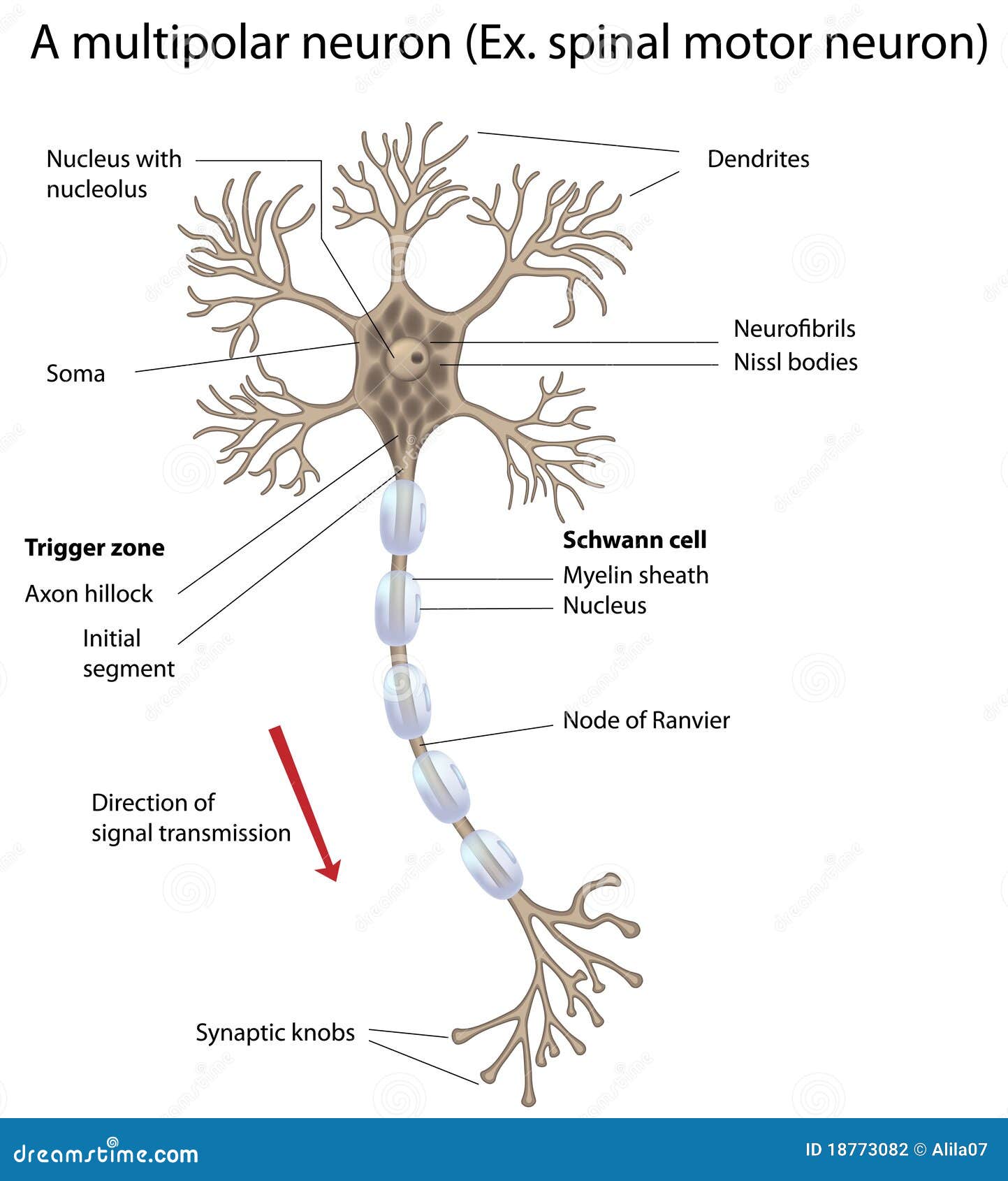
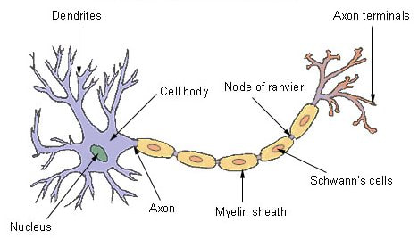
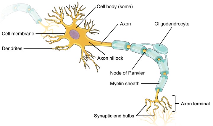
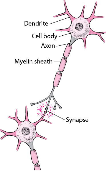
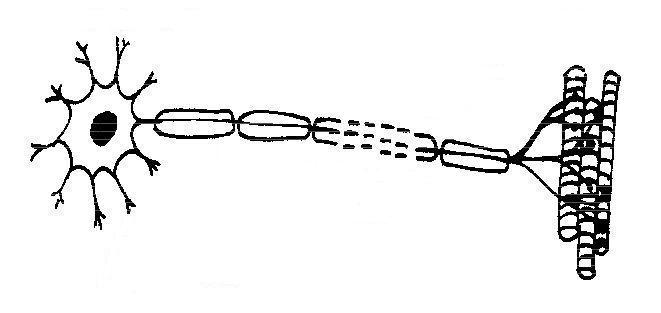
43 nerve cell drawing and label
Plant Cell Model - Labeled & Unlabeled Halves - Home Science Tools One half is labeled with plant cell parts: mitochondria, smooth and rough ER, chloroplasts, Golgi apparatus, vacuole, ribosomes, cytoplasm, cell wall, and nucleus with nucleolus and chromatin. The other half has the same parts, only unlabeled for testing. Includes booklet with facts about cells, a diagram to label, information on photosynthesis ... EOF
Cross sectional anatomy | Kenhub Cross-sections are two-dimensional, axial views of gross anatomical structures seen in transverse planes. They are obtained by taking imaginary slices perpendicular to the main axis of organs, vessels, nerves, bones, soft tissue, or even the entire human body. Cross-sections provide the perception of 'depth', creating three-dimensional ...
/548299803-56a796543df78cf7729765c8.jpg)
Nerve cell drawing and label
Draw A Sheep And Label - Warehouse of Ideas Draw a label diagram of a sheep keywords: Source: . The neuron sends electrical impulses from its cell body through the axon to target cells. Draw a label diagram of a sheep the items of militaria shown below can be viewed in our on line shop complete with full descriptions photographs and prices b e f slip on shoulder title a ... The 9 parts of a neuron (and their functions) - medical - 2022 Nissl substance. 7. Ranvier's nodules. 8. Synaptic buttons. 9. Axonal cone. Bibliographic references. Neurons are a type of cells in our body that are incredibly specialized on a morphological level. and physiological in fulfilling an essential function: transmitting information throughout the body. High-speed camera captures signals traveling through nerve cells Some nerve signals are even faster, approaching speeds of 300 miles per hour. Now, scientists at Caltech have developed a new ultrafast camera that can record footage of these impulses as they ...
Nerve cell drawing and label. Team discovers second stem cell type in mouse brain In the brain of adult mammals, neural stem cells ensure that new nerve cells (neurons), are constantly formed. This process, known as adult neurogenesis, helps mice maintain their sense of smell ... Somatic Cells - Genome.gov Somatic cells. All organisms that are alive are made of one or more cells that are called somatic cells. In humans, somatic cells are diploid, meaning they contain two sets of chromosomes, one set inherited from each parent. Somatic mutations can impact the individual carrying the mutation, but cannot be passed on and have no effect on the ... Spinal Cord Cross Section | New Health Advisor The nerve bundle has a cushion of cerebrospinal fluid between it and the meninges. The end of the spinal cord is called the cauda equine because it looks like a horse's tail with its cascade of nerves. The Structure. Exiting through a big hole at the bottom of the skull, the spinal cord is covered by the vertebral column that protects it. The Survival of Nerve Cells, As Well As their Performance of Some ... Reading Passage Question: The survival of nerve cells, as well as their performance of some specialized functions, is regulated by chemicals known as neurotrophic factors, which are produced in the bodies of animals, (5) including humans. Rita Levi-Montalcini's discovery in the 1950s of the first of these agents, a hormonelike substance now ...
Cell Cycle - Genome.gov Cell cycle is the name we give the process through which cells replicate and make two new cells. Cell cycle has different stages called G1, S, G2, and M. G1 is the stage where the cell is preparing to divide. To do this, it then moves into the S phase where the cell copies all the DNA. So, S stands for DNA synthesis. Neuromuscular Junction Structure and Functions - New Health Advisor The synapse or connection between a motor neuron and a skeletal muscle is known as neuromuscular junction. Communication happens between the neuron and muscle via nerve cells. Due to this communication or transmission of signal, the muscle is able to contract or relax. It is the most widely studied synapse and it is comparatively easier to ... Second stem cell type discovered in mouse brain In the brain of adult mammals neural stem cells ensure that new nerve cells, i.e. neurons, are constantly formed. This process, known as adult neurogenesis, helps mice maintain their sense of ... Cranial nerves: Anatomy, names, functions and mnemonics | Kenhub Anatomy. Cranial nerves are the 12 nerves of the peripheral nervous system that emerge from the foramina and fissures of the cranium.Their numerical order (1-12) is determined by their skull exit location (rostral to caudal). All cranial nerves originate from nuclei in the brain.Two originate from the forebrain (Olfactory and Optic), one has a nucleus in the spinal cord (Accessory) while the ...
High-speed camera captures signals traveling through nerve cells. Some nerve signals are even faster, approaching speeds of 300 miles per hour. Now, scientists at Caltech have developed a new ultrafast camera that can record footage of these impulses as they travel through nerve cells. The camera can also capture video of other ultrafast phenomena, like the propagation of electromagnetic pulses in electronics. High-speed camera captures signals traveling through nerve cells ... Some nerve signals are even faster, approaching speeds of 300 miles per hour. Now, scientists at Caltech have developed a new ultrafast camera that can record footage of these impulses as they travel through nerve cells. The camera can also capture video of other ultrafast phenomena, like the propagation of electromagnetic pulses in electronics. Mitochondrion - Wikipedia A mitochondrion (/ ˌ m aɪ t ə ˈ k ɒ n d r i ə n /; pl. mitochondria) is a double-membrane-bound organelle found in most eukaryotic organisms. Mitochondria use aerobic respiration to generate most of the cell's supply of adenosine triphosphate (ATP), which is subsequently used throughout the cell as a source of chemical energy. They were discovered by Albert von Kölliker in 1857 in the ... nervous system | Definition, Function, Structure, & Facts A nervous system can be defined as an organized group of cells, called neurons, specialized for the conduction of an impulse—an excited state—from a sensory receptor through a nerve network to an effector, the site at which the response occurs. Organisms that possess a nervous system are capable of much more complex behaviour than are ...
High-speed camera captures signals traveling through nerve cells Some nerve signals are even faster, approaching speeds of 300 miles per hour. Now, scientists at Caltech have developed a new ultrafast camera that can record footage of these impulses as they ...
The 9 parts of a neuron (and their functions) - medical - 2022 Nissl substance. 7. Ranvier's nodules. 8. Synaptic buttons. 9. Axonal cone. Bibliographic references. Neurons are a type of cells in our body that are incredibly specialized on a morphological level. and physiological in fulfilling an essential function: transmitting information throughout the body.
Draw A Sheep And Label - Warehouse of Ideas Draw a label diagram of a sheep keywords: Source: . The neuron sends electrical impulses from its cell body through the axon to target cells. Draw a label diagram of a sheep the items of militaria shown below can be viewed in our on line shop complete with full descriptions photographs and prices b e f slip on shoulder title a ...
















Post a Comment for "43 nerve cell drawing and label"