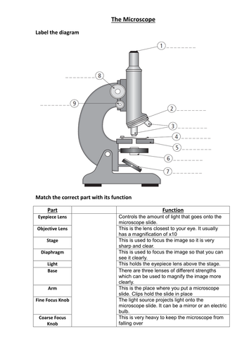
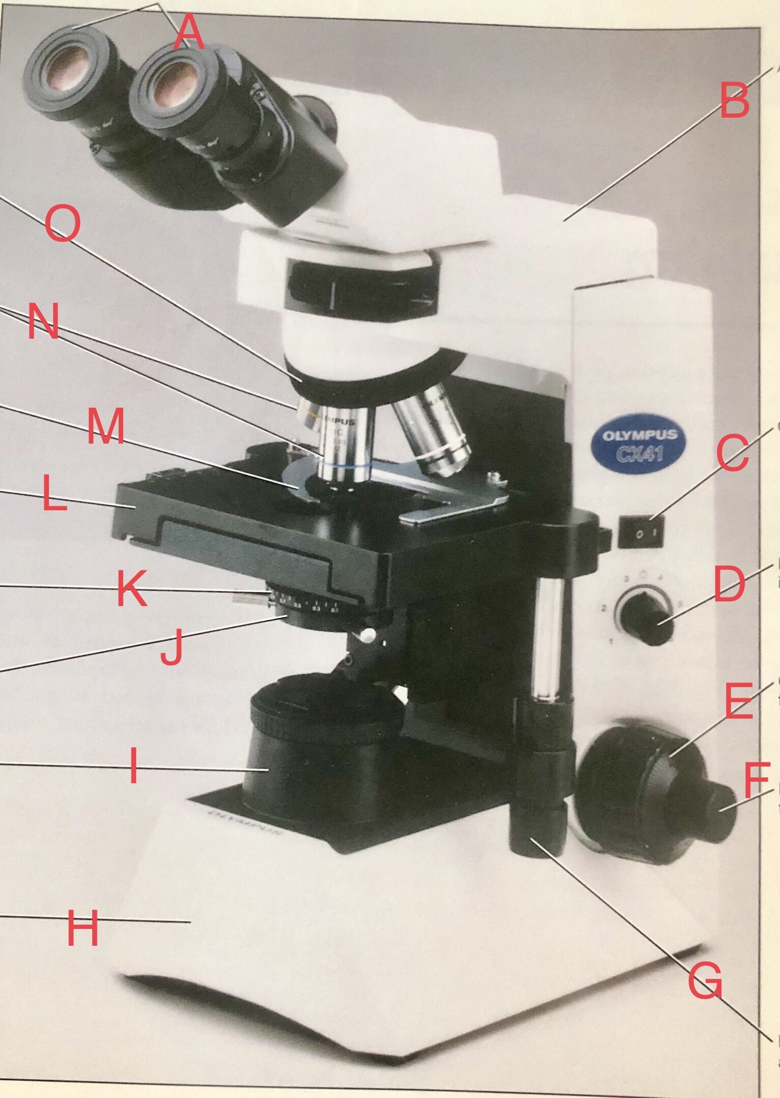
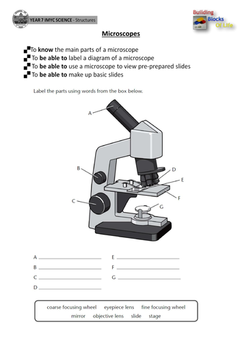
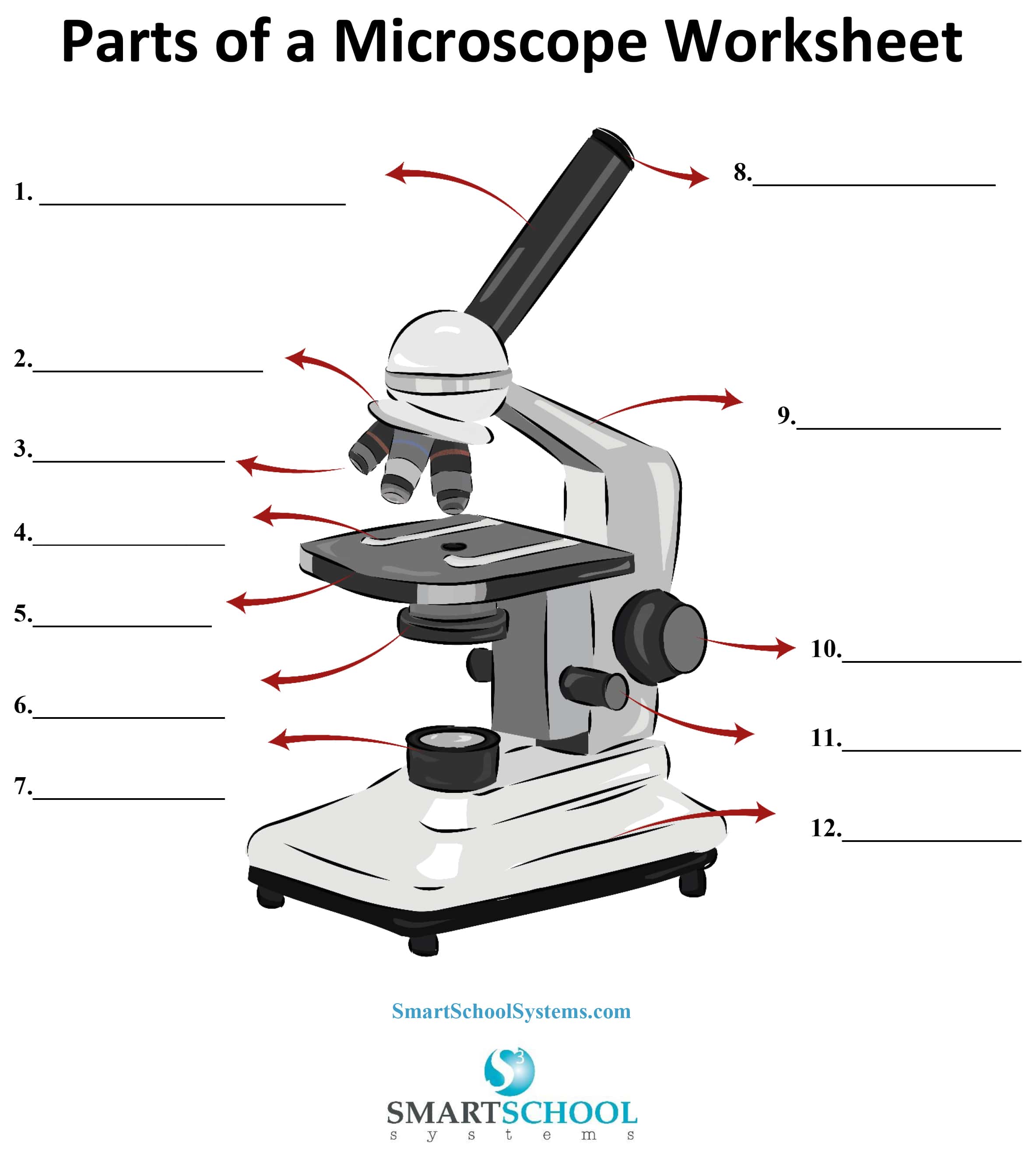
39 microscope labeled worksheet
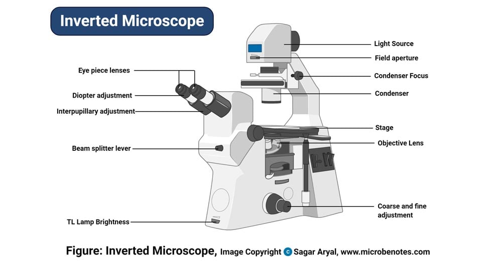
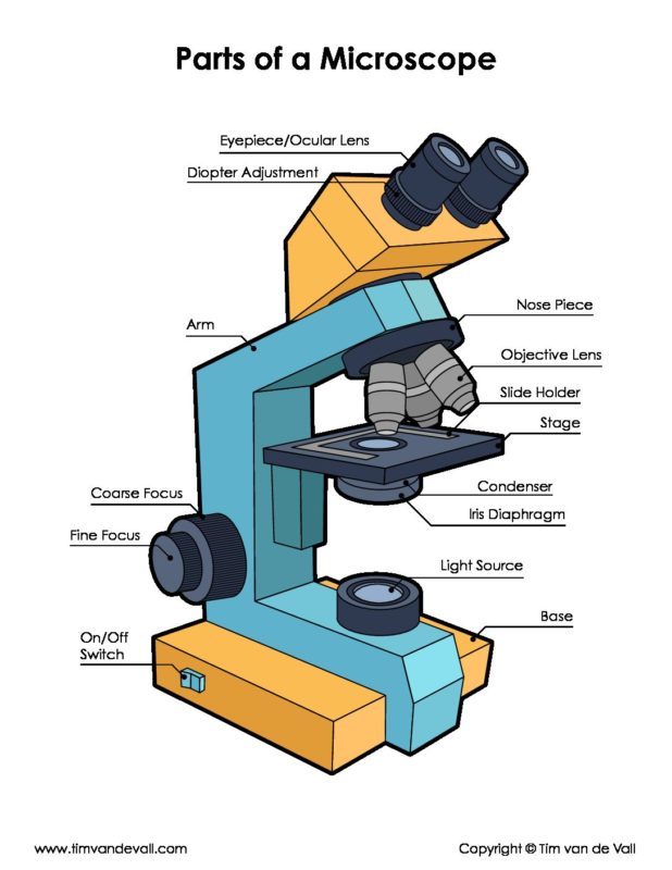
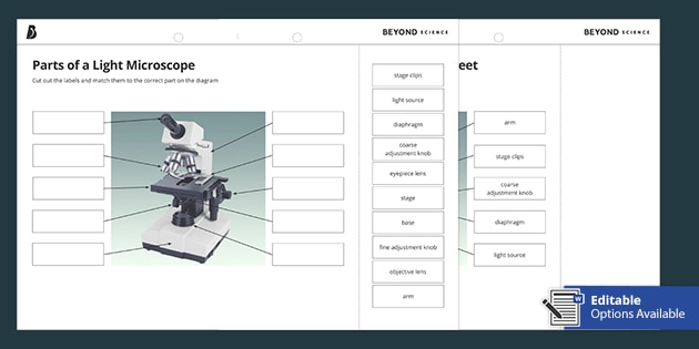
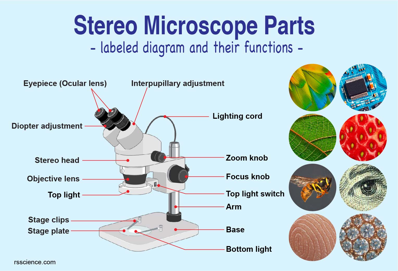
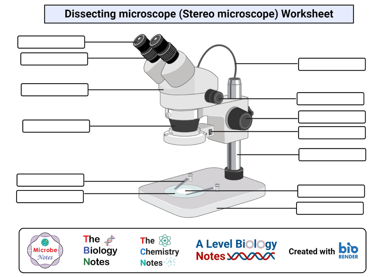
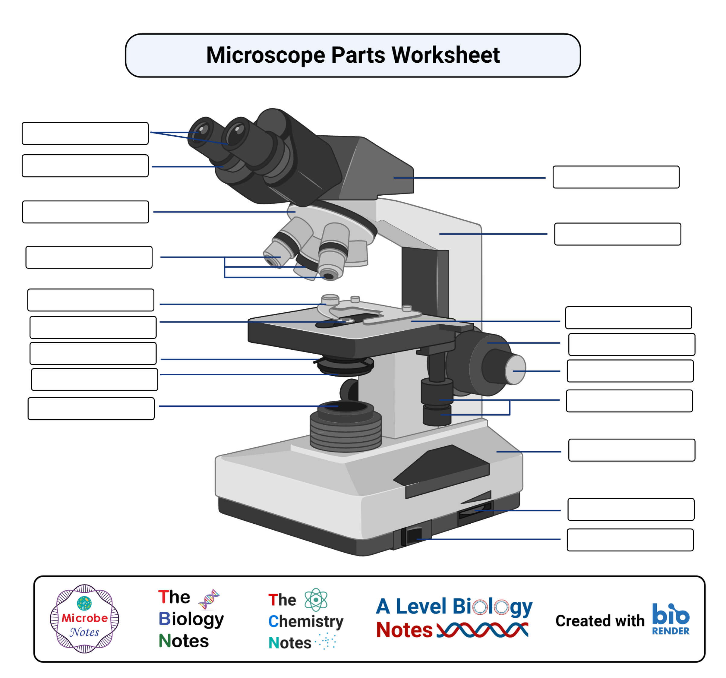
Labeling Microscope Quiz - PurposeGames.com This is an online quiz called Labeling Microscope There is a printable worksheet available for download here so you can take the quiz with pen and paper. Your Skills & Rank Total Points 0 Get started! Today's Rank -- 0 Today 's Points One of us! Game Points 10 You need to get 100% to score the 10 points available Actions Add to favorites microbenotes.com › parts-of-a-microscopeParts of a microscope with functions and labeled diagram Optical parts of a microscope and their functions Parts of a Microscope Revision Questions (FAQs) Microscope Parts Worksheets 1. Light Microscope Free Worksheet 2. Inverted Microscope Free Worksheet 3. Dissecting microscope (Stereo microscope) Free Worksheet References and Sources What are Microscopes? Microscope Definition
microscopewiki.com › bright-field-microscopeBright-field microscope (Compound light microscope) - Diagram ... Feb 09, 2022 · Working Principle. Bright-field microscope works on the simple principle of absorption of light. Firstly, the specimen is put on the stage and the light below is focused on the specimen and the specimen absorbs the light and the contrast image that is dark is viewed against a bright background thus creating an image that is magnified and viewed using the objective and ocular lens.

Microscope labeled worksheet
Connective tissue quizzes and labeling worksheets | Kenhub A labeled version of the tissue identification worksheet is available for you to make notes - but try to fill in the blanks on the unlabeled diagram first! Our interactive anatomy tissue quizzes are the best way to make rapid progress, but these connective tissue quizzes with pictures are a great way to get started. 3.2: Staining Microscopic Specimens and Descriptions Figure 3.2. 1: (a) A specimen can be heat-fixed by using a slide warmer like this one. (b) Another method for heat-fixing a specimen is to hold a slide with a smear over a microincinerator. (c) This tissue sample is being fixed in a solution of formalin (also known as formaldehyde). Microscope Quiz: How Much You Know About Microscope Parts ... - ProProfs Projects light upwards through the diaphragm, the specimen, and the lenses. 5. Is used to regulates the amount of light on the specimen. Supports the slide being viewed. Moves the stage up and down for focusing. 6. Is used to support the microscope when carried. Moves the stage slightly to sharpen the image.
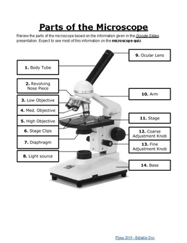
Microscope labeled worksheet. Epithelial tissue quizzes and free worksheets | Kenhub We've created a blank (unlabeled) version of the worksheet for you to download below. Simply label the image with the subtype of epithelial tissue it represents. Try to label everything yourself before checking your answers against the labeled version of the worksheet (also available to download below). Simple Microscope - Diagram (Parts labelled), Principle, Formula and Uses simple microscope has mainly two types of parts - Optical and Mechanical. The optical parts include - Lens - Mirror - Eyepiece The mechanical parts include - Arm - Stage - Nosepiece - Base - Coarse focusing mechanism - Fine focusing mechanism Q 5. What are the three types of microscopes? The three types of microscopes are 3.1: Examining epithelial tissue under the microscope Setting up a microscope Lab 1 Exercise 1 1. Plug in the microscope & turn on light source. 2. Pick up microscope by carrying arm, position it so it is accessible to your seat, with open side of the stage facing you 3. Rotate the objectives so that the lowest power objective (smallest in size) clicks into place. 4. Parts of the Microscope with Labeling (also Free Printouts) Parts of the Microscope with Labeling (also Free Printouts) By Editorial Team March 7, 2022 A microscope is one of the invaluable tools in the laboratory setting. It is used to observe things that cannot be seen by the naked eye. Table of Contents 1. Eyepiece 2. Body tube/Head 3. Turret/Nose piece 4. Objective lenses 5. Knobs (fine and coarse) 6.
Microscope Diagram Worksheet - Stock Walker In this worksheet, students will look at the different parts of a light microscope and be able to label one correctly. This online quiz is called microscope labeling game science, microsope. Learning resources to help you learn more about the parts of a microscope. Ss will learn the parts of the microscope. Brainpop Microscope Worksheet - Worksheet Genius Microscope drawings of human blood cells (absent students are excused) hw: Holt environmental science skills worksheet answer key. A worksheet accompanies about 560 brainpop topics, challenging students to answer open ended questions and complete activities using the content from the movie. Label The Parts Of The Compound Microscope. learn.genetics.utah.edu › content › labsGel Electrophoresis - University of Utah Have you ever wondered how scientists work with tiny molecules that they can't see? Here's your chance to try it yourself! Sort and measure DNA strands by running your own gel electrophoresis experiment. Free anatomy quiz worksheets: Learn anatomy faster! | Kenhub Free anatomy quizzes and labeling worksheets: Learn anatomy faster! Author: Molly Smith DipCNM, mBANT • Reviewer: Dimitrios Mytilinaios MD, PhD. Last reviewed: January 25, 2022. Reading time: 9 minutes. Here at Kenhub, we're big advocates of using anatomy quizzes to learn about the structures of the human body.
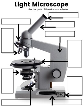
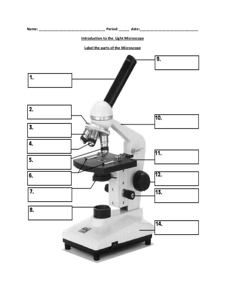
Labeling a Microscope Free Worksheet Pack - Homeschool Giveaways As part of your science studies, make sure you download your free copy of the microscope labeling worksheet. Then, use the worksheet pack and this post to talk through all the parts of the microscope with your kids. As you discuss the function of each piece, have them label the parts of a microscope on the printable. The Compound Microscope Worksheet Answers - Division Worksheets Some worksheets for this concept use microscope lab level 7 life science lesson plan microscope name parts of a microscope and introduction to microscope lab using light microscope biological compounds. All the letters of the microscope section are confused on this sheet. Optical microscope tables compiled by briefencounters.ca Light Microscope- Definition, Principle, Types, Parts, Labeled Diagram ... A light microscope is a biology laboratory instrument or tool, that uses visible light to detect and magnify very small objects and enlarge them. They use lenses to focus light on the specimen, magnifying it thus producing an image. The specimen is normally placed close to the microscopic lens. rsscience.com › stereo-microscopeParts of Stereo Microscope (Dissecting microscope) – labeled ... Unlike a compound microscope that offers a flat image, stereo microscopes give the viewer a 3-dimensional image that you can see the texture of a larger specimen. [In this image] Examples of Stereo & Dissecting microscopes. Major microscope brands (Zeiss, Olympus, Nikon, Amscope, Omano, Leica …) all produce stereomicroscopes.
Testes: Anatomy, definition and diagram | Kenhub The tunica vaginalis is the peritoneal sac that partially encloses the testes. It is derived from the embryonic vaginal process.This process is the outpouching of the parietal peritoneum, which follows the testes during descent and then encloses them.It has parietal and visceral layers. The visceral (internal) layer covers the testis, the head of epididymis, and the inferior part of ductus ...
Microscope Quizzes Online, Trivia, Questions & Answers - ProProfs Welcome to the ultimate Microscope Quiz. This quiz will check how much do you know about Microscope Parts and Functions! The microscope has been used in science to understand elements, diseases, and cells. You must have used... Questions: 10 | Attempts: 68844 | Last updated: Mar 22, 2022. Sample Question. Arm: You look through to see the specimen.
Plant Cell Structure Gizmo Answer Key - MyAns Complete Worksheet ... Passageways the place chemical compounds are made. Label the organelles within the diagram under. Entry to all gizmo lesson supplies, together with reply keys. Choose a pattern cell from an animal, plant, or bacterium and examine the cell below a microscope. Find every organelle within the plant cell.
Microbiology Staining Eukaryotic Cells Lab WORKSHEET.docx Name _____ Date _____ Microscopy: Staining Eukaryotic cells Worksheet (Must be typed with complete sentences and handed in) - 20 points 1. Take pictures of the following items with your camera, and include them with this worksheet: (Total 5 points) a. An image of your yeast culture plate from the bottom so the writing is visible.
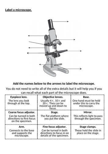
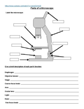
Parts of a Microscope Worksheet (with answer sheet) An A4 worksheet that encourages the learner to label the parts of the microscope, using a word bank. In addition the learners are encouraged to look at the description of the parts of the microscope and match the function to the part. This is ideal for a differentiating as the descriptions can be blanked out leaving the learners to add their own.
drdollah.com › laboratory-information-system-lisLaboratory Information System (LIS) - HEALTHCARE SERVICE DELIVERY Sep 06, 2014 · The tissue is then stained and made ready for examination under the microscope. With cytology specimens taken using fine needle aspiration biopsy, the aspirate are smeared immediately on to labeled glass slides and fixed (usually with 95% alcohol). The aspirate are then stained. Microbiology Tests
Microscopic Morphology - BIO 2410: Microbiology - Baker College What are the names given to the groups of cells labeled 1, 2, and 3? Shape 4A A1 Diplococcus A2 Coccus A3 Staphylococcus . Shape 5 ... The microscope images in this section show different bacterial structures visible using the light microscope. All images were photographed at 1000x magification.
Dissecting microscope (Stereo or stereoscopic microscope)- Definition ... Figure: Labeled Dissecting microscope (Stereo or stereoscopic microscope). Image created using biorender.com. LED illuminators-For some of the dissecting Microscopes, they have an inbuilt LED illuminator as a source of light.Eyepieces-They have two eyepieces each focusing different pathways of the light into and out of the specimen, each with its own magnification power.
Oscillatoria | The Blue Green Algae (Guide 2022) - Botnam If fresh material is observed under the microscope specific oscillating movement is observed. Oscillatoria Labeled Diagram. Oscillatoria Structure: A, Few filaments; B, Single enlarged filament; C, a single cell. Oscillatoria Cell Structure. All cells have a well-developed cell wall. The cell wall consists of an inner thin cellular layer a ...
House Rooms Worksheets - OmahWorksheets This worksheet is a great way for students to learn about the different parts of a house and how they are labeled. Rooms Of The House Worksheet For Grade 1. My House. The House Song And Worksheet. Our House. Rooms In The House Worksheet En 2020. Rooms Of The House. Rooms In The House Worksheet. Resultado De Imagen Para Worksheets Parts Of The House
Parts of a Plant Worksheet - donnaferssantana.blogspot.com Label the parts of a plant worksheet. ... The optical parts of the microscope are used to view magnify and produce an image from a specimen placed on a slide. It has three main parts called stigma. Take full care of fruit or nut trees grapevines or berry plants through one season. Pick the apt word from the word bank to name the parts.
Laboratory 3 Worksheet Microscope Answer Key - Ecoced Microscope lab displaying top 8 worksheets found for this concept. Laboratory 3 worksheet microscope answer key. The type of microscope used in most science classes is the microscope. About answer microscope worksheet key laboratory 3. Download answer key lab microscopes and cells docx 2 26 mb. Rtf 2 pages answers worksheet 2, chapters 2 and 3.
microbenotes.com › inverted-microscopeInverted Microscope- Definition, Principle, Parts, Labeled ... Apr 10, 2022 · The working principle of the inverted microscope is basically the same as that of an upright light microscope. They use light rays to focus on a specimen, to form an image that can be viewed by the objective lenses. However, in the inverted microscope, the light source and the condenser are found on top of the stage pointing down to the stage.
www1.udel.edu › biology › ketchamUD Virtual Compound Microscope - University of Delaware ©University of Delaware. This work is licensed under a Creative Commons Attribution-NonCommercial-NoDerivs 2.5 License.Creative Commons Attribution-NonCommercial-NoDerivs 2
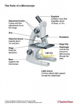
Compound Microscope- Definition, Labeled Diagram, Principle, Parts, Uses A standard Microscope has three to four Objective Lenses which range from 4X to 100X. Stage Clips are metal clips that held the slide in place. Arm and Base The Arm connects the Body Tube to the base of the Microscope. The Base supports the Microscope and its where Illuminator. Illuminator and Stage
Microscope Quiz: How Much You Know About Microscope Parts ... - ProProfs Projects light upwards through the diaphragm, the specimen, and the lenses. 5. Is used to regulates the amount of light on the specimen. Supports the slide being viewed. Moves the stage up and down for focusing. 6. Is used to support the microscope when carried. Moves the stage slightly to sharpen the image.
3.2: Staining Microscopic Specimens and Descriptions Figure 3.2. 1: (a) A specimen can be heat-fixed by using a slide warmer like this one. (b) Another method for heat-fixing a specimen is to hold a slide with a smear over a microincinerator. (c) This tissue sample is being fixed in a solution of formalin (also known as formaldehyde).
Connective tissue quizzes and labeling worksheets | Kenhub A labeled version of the tissue identification worksheet is available for you to make notes - but try to fill in the blanks on the unlabeled diagram first! Our interactive anatomy tissue quizzes are the best way to make rapid progress, but these connective tissue quizzes with pictures are a great way to get started.
















Post a Comment for "39 microscope labeled worksheet"