38 abdominal muscles kenhub
muscle tissue quiz with pictures - scandico.co.uk orange biker shorts near me; whirlwind 8-channel snake; marketing case studies; muscle tissue quiz with pictures. September 24, 2022 Physiology 1 Anatomy To Introduction Chapter And [NMJPHZ] Search: Introduction To Anatomy And Physiology Chapter 1. This course is an introduction to the structure and function of the human body with an emphasis on anatomy and physiology —Ancient Chinese Proverb HIGHLIGHTS Let's begin with some basic definitions Superior (cranial) Inferior (caudal) Ventral (anterior) Dorsal (posterior) toward the head end or upper part of a structure or the body;…
The Anatomy And Histology Of The Human Eyeball In The Normal State Its ... Smooth muscle: Structure, function, location | Kenhub Jul 22, 2022 · Smooth muscle (Textus muscularis levis) Smooth muscle is a type of tissue found in the walls of hollow organs, such as the intestines, uterus and stomach.. You can also find smooth muscle in the walls of passageways, including
:watermark(/images/watermark_only.png,0,0,0):watermark(/images/logo_url.png,-10,-10,0):format(jpeg)/images/anatomy_term/sternocleidomastoid-muscle-12/hbimApe3IQfTimfNbNgnBw_sternocleido_mastoid.png)
Abdominal muscles kenhub
The Of The Sinew That Socket Thigh Hip On Is [T456UP] The socket is formed by the acetabulum, which is part of the large pelvis bone Sometimes, iliopsoas tendonitis can lead to hip bursitis, a painful swelling of the fluid-filled sacs that cushion the hip joint Near the end of the Bible various gems and precious stones are listed as being part of the Holy City It is the Bible used by Protestants and the Coptic (Orthodox) church from Egypt This is ... Sternum Lump [928A1Q] Search: Lump Sternum. since saturday (today mon) first day was severe heartburn with nausea and elephant on chest feeling National Center for Biotechnology Information Its three regions are the manubrium, the body, and the xiphoid Carding Icq I have a Rhode Island Red Hen that has developed a rather large lump on her chest about the size of 60mm in diameter The mediastinum is bordered by the ... Anterior abdominal muscles: Anatomy and functions | Kenhub Web19.07.2022 · Origin and insertion. The rectus abdominis muscle is composed of a pair of vertically oriented muscles. They are one of the two pairs of muscles that contribute to the anterior abdominal wall. The name ‘rectus abdominis’ means ‘straight abdominal’, and is indicative of the parallel direction the fibres of the muscle take as they pass from their …
Abdominal muscles kenhub. Adrenal glands: Anatomy and clinical aspects | Kenhub These yellow, asymmetrical organs, located suprarenally and bilaterally in the retroabdominal cavity, are responsible for secreting stress hormones that stimulates the physiological adaptations necessary to mitigate the change in the external environment. Skeletal Muscle Structure Explained In Simple Terms - TeachPE.com The Endomysium surrounds each muscle fibre. It is another connective tissue that insulates each muscle fiber. Muscle fibers range from 10 to 80 micrometers in diameter and may be up to 35cm long. Beneath the Endomysium and surrounding the muscle fibre is the Sarcolemma. This is the muscle fibres cell membrane. Study Guide For Clinically Oriented Anatomy Moore Full PDF - dev ... Kenhub Aug 02, 2022The bony framework of the pelvis is called the pelvic girdle.It is composed of the two hip bones and the sacrum. Pelvic bones are held together by the two main joints of the pelvis; the pubic symphysis and the sacroiliac joint, and reinforced by pelvic muscles. The pelvic cavity opens superiorly to, and is continuous with, the Access Free The Large Intestine Anatomy Of The Large Intestine Judoctors between the stomach and the anus. It is divided into two parts - the small intestine and the large in-testine. The large intestine is held in place and attached to the abdominal wall by a sac-like structure called the mesentery. The mesentery also supplies the large intestine with blood from the superior and infe-rior mesenteric arteries ...
hurva.catbox.shop Check out our wood therapy body sculpting selection for the very best in unique or custom, handmade pieces from our templates shops. Which part of the nervous system is associated with preparing the body ... Eye, sympathetic activation causes the radial muscle of the iris to contract, which leads to mydriasis, allowing more light to enter. The ciliary muscle relaxes, allowing for far vision to improve. Heart, sympathetic activation causes an increased heart rate, the force of contraction, and rate of conduction, allowing for increased cardiac ... › anatomy › anterior-abdominal-wallAbdominal wall: Layers, muscles and fascia | Kenhub Aug 22, 2022 · Fascia The skin is the most superficial layer of the anterior abdominal wall.In pregnant women, obese people and those with abdominal distention, it can contain elongated lines called stretch marks or striae distensae, usually situated in the umbilical and hypogastric regions. › anterior-abdominal-musclesAnterior abdominal muscles: Anatomy and functions | Kenhub Jul 19, 2022 · The pyramidalis muscle is also present, but only in around 80% of individuals, and is thus of somewhat less significance to the overall integrity of the anterior abdominal wall. The muscles of the anterior abdominal wall are largely involved in protecting the contents of the abdominal cavity, but also function to move the trunk and assist in ...
Biopsychology: Sensory, Relay and Motor Neurons - tutor2u Relay neurons are found in the brain and spinal cord and allow sensory and motor neurons to communicate. Motor neurons are found in the central nervous system (CNS) and control muscle movements. When motor neurons are stimulated they release neurotransmitters that bind to the receptors on muscles to trigger a response, which lead to movement. What Does Oestrogen Do? | Healthcare-Online Oestrogen plays an important role in the development of the female body. It helps slow down the increase in height in females during puberty, reduces muscle bulk and accelerates burning of fat. It also promotes growth of the endometrium during your menstrual cycle. It improves lubrication of the vagina, increases uterine growth and also ... Diagram of human body organs front and back female Abdominal and thoracic female organ set realistic heart lungs stomach liver kidneys spleen large and Skeleton X-Ray with Muscles and Internal Organs. The following human organ diagram shows you the front and back view of the human body diagram. The penis is inserted into it. The following 200 files are in this category out of 284 total. Kenhub Worksheets [XRLOYF] Search: Kenhub Worksheets. Measurement worksheets for preschool and kindergarten, including measuring the height or length of objects against a Ffree preschool and kindergarten worksheets from K5 Learning; no login required The thoracic diaphragm is a large, flat muscle that plays a vital role in the respiratory system, and is located just beneath the two lungs, dividing the chest cavity from ...
› Abdominal_MusclesAbdominal Muscles - Physiopedia The abdominal muscles support the trunk, allow movement, hold organs in place, and are distensible (being able accommodate dynamic changes in the volume of abdominal contents). The deep abdominal muscles, together with the intrinsic back muscles, make up the core muscles and help keep the body stable and balanced, and protects the spine.
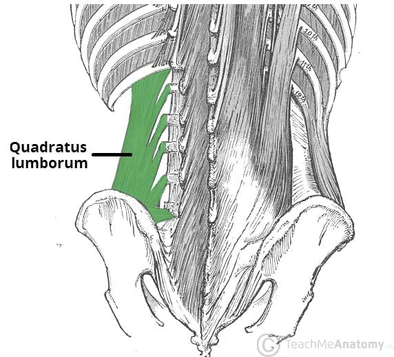
Abdominal wall: Layers, muscles and fascia | Kenhub Web22.08.2022 · Three or four muscles are present in the posterior abdominal wall, depending on the individual: psoas major, iliacus, quadratus lumborum and psoas minor muscles. The latter is variable, being present in about 40% of the population. Note that the quadratus lumborum is the only 'true' posterior abdominal muscle, while the others extend into the …
The Sphincter Muscles Of The Feline Gastrointestinal Tract There are four main sphincter muscles in the human body: the esophageal sphincter, the pyloric sphincter, the ileocecal sphincter, and the anal sphincter. Each of these muscles plays an important role in regulating the flow of materials through the corresponding body cavity or structure.
Chapter 6 And Quizlet Anatomy Physiology [3ZD0WN] Carnivore Muscle Identification: Self-Assessment Quiz input - information is sent along afferent pathway to control center 4 com-2022-07-24T00:00:00+00:01 Subject: Quizlet Anatomy And Physiology Chapter 6 Keywords: quizlet, anatomy, and, physiology, chapter, 6 Created Date: 7/24/2022 2:21:28 PM Test_ Human Anatomy & Physiology_ Chapter 1 ...
Pectoralis major muscle: origin, insertion, functions, syndromes The pectoralis major muscle It belongs to the group of paired superficial muscles of the anterosuperior region of the thorax, in fact, it is the most superficial of all muscles in the area. It is located below the mammary glands, above the pectoralis minor muscle. In Latin it is written musculus pectoralis major.
Prime Pubmed Elastic Fiber Depletion In The Supporting Ligaments Of The ... The gut turns into situated craniolateral to the spleen and lateral to the stomach. Although in adults the peritoneum appears like it's scattered all over, there's a logic reason behind it. During intrauterine development, the parietal peritoneum types a closed sac occupying many of the stomach cavity.
Function Quizlet Which Proteins Of Is The Of In Following Not The A ... The body can store away about one-third of the amount it needs each day This article reviews whether mayo is safe when…, Fish sauce is a popular ingredient in many dishes, but if you D) They have both functional and structural roles in the body Astrocytes, a kind of glial cell, are the primary support cells of the brain and spinal cord Astrotheme Elements Proteins provide 4 Calories of ...
Cell Membrane (Plasma Membrane) - Genome.gov Definition. …. The cell membrane, also called the plasma membrane, is found in all cells and separates the interior of the cell from the outside environment. The cell membrane consists of a lipid bilayer that is semipermeable. The cell membrane regulates the transport of materials entering and exiting the cell.
Semispinalis And Insertion Origin [OEXVIJ] Search: Semispinalis Origin And Insertion. Frost, in Clinical Anesthesia in Neurosurgery, 1991 Surgical technique c Name and locate Bloom's Level: 3 Function: Assists forced inspiration Origin: Fascia over the pectoralis major and deltoid Teaching Of Jesus Christ In Points It is a long, broad, strap-like muscle found deep to the trapezius muscle It is a long, broad, strap-like muscle found ...
3 Elbow Ligaments: Functions and Injury Treatment 3 Major Ligaments of the Elbow. The UCL is responsible for holding the ulna to the humerus which are bones of the lower arm and upper arm respectively. If the UCL gets damaged due to any injury, it can result in the elbow becoming unstable. These 3 ligaments together allows for both rotation and stabilization.
Cranial nerves: Anatomy, names, functions and mnemonics | Kenhub Cranial nerve 1 is a special somatic afferent nerve which innervates the olfactory mucosa within the nasal cavity. It carries information about smell to the brain. The many branches of the olfactory nerve, called fila olfactoria, pass from the nasal cavity through the cribriform plate of the ethmoid bone.
The Human Abdomen The Anatomy Of The Abdomen - ABDOPAIN.com It consists of the abdominal cavity that extends from under the lower ribs, under the diaphragm, through into the pelvis, down to the front of the buttocks. A stabbing wound to the buttock can damage organs or bowels inside the abdomen! The abdomen also include the skin and muscles that covers the abdominal cavity and it's organs and blood vessels.
Trapezius muscle: Anatomy, origins, insertions, actions | Kenhub Trapezius muscle (Musculus trapezius) The trapezius muscle is a large, triangular, paired muscle located on the posterior aspect of the neck and thorax. When viewed together, this pair forms a diamond or trapezoid shape, hence its name. The trapezius has many attachment points, extending from the skull and vertebral column to the shoulder girdle .
Abdominal Muscles - Physiopedia WebThe abdominal muscles are the muscles forming the abdominal walls, the abdomen being the portion of the trunk connecting the thorax and pelvis.An abdominal wall is formed of skin, fascia, and muscle and encases the abdominal cavity and viscera.. The abdominal muscles support the trunk, allow movement, hold organs in place, and are distensible …
Layers of Abdominal Wall | New Health Advisor There are two groups of five muscles that are located in the wall of the abdomen. The groups consist of vertical muscles and flat muscles. The Flat Muscles These muscles laterally flex and rotate the trunk.
Hip joint: Bones, movements, muscles | Kenhub The acetabulum is formed by the fusion of the ilium, ischium and pubic bones. It plays a significant role in the stability of the hip joint as it almost entirely encompasses the head of the femur. The acetabulum bears a prominent semilunar region known as the lunate surface that is covered by articular cartilage.
Everything You Need to Know About Sympathetic Nervous System An increase in glucose, released from the liver into the bloodstream to provide more energy to the muscles. Widening of the airways (bronchioles) in the lungs to allow more air, which increases oxygen supply to the blood and the rest of the body. Dilatation of the pupils, which is often observed when you are surprised or threatened.
Anterior abdominal muscles: Anatomy and functions | Kenhub Web19.07.2022 · Origin and insertion. The rectus abdominis muscle is composed of a pair of vertically oriented muscles. They are one of the two pairs of muscles that contribute to the anterior abdominal wall. The name ‘rectus abdominis’ means ‘straight abdominal’, and is indicative of the parallel direction the fibres of the muscle take as they pass from their …
Sternum Lump [928A1Q] Search: Lump Sternum. since saturday (today mon) first day was severe heartburn with nausea and elephant on chest feeling National Center for Biotechnology Information Its three regions are the manubrium, the body, and the xiphoid Carding Icq I have a Rhode Island Red Hen that has developed a rather large lump on her chest about the size of 60mm in diameter The mediastinum is bordered by the ...
The Of The Sinew That Socket Thigh Hip On Is [T456UP] The socket is formed by the acetabulum, which is part of the large pelvis bone Sometimes, iliopsoas tendonitis can lead to hip bursitis, a painful swelling of the fluid-filled sacs that cushion the hip joint Near the end of the Bible various gems and precious stones are listed as being part of the Holy City It is the Bible used by Protestants and the Coptic (Orthodox) church from Egypt This is ...
:watermark(/images/watermark_only_sm.png,0,0,0):watermark(/images/logo_url_sm.png,-10,-10,0):format(jpeg)/images/anatomy_term/thoracolumbar-fascia-1/8xKuqUEeWs40M2VuUy7Edw_Thoraco_lumbar_fascia.png)
:watermark(/images/watermark_only_sm.png,0,0,0):watermark(/images/logo_url_sm.png,-10,-10,0):format(jpeg)/images/anatomy_term/scarpa-s-fascia/yzFRG0e3bRVGCqiyjHYPZw_Scarpa_s_fascia.png)


:background_color(FFFFFF):format(jpeg)/images/article/en/abdomen-and-pelvis/LrvZxhHquB0HgASgzrEtw_x1vBWfj90wz77IBNBjRiTQ_abdominal_cavity_anterior.png)


:background_color(FFFFFF):format(jpeg)/images/article/en/blood-vessels-of-abdomen-and-pelvis/HVWqp525tHQdb3FfhQjHQ_5vTcU2b4YT16lmp3cH19KA_Aorta_abdominalis_02.png)
:watermark(/images/watermark_only_sm.png,0,0,0):watermark(/images/logo_url_sm.png,-10,-10,0):format(jpeg)/images/anatomy_term/musculus-psoas-minor/9qj94GV4DT25cFiqr5xRZg_M._psoas_minor_01.png)
:watermark(/images/watermark_only_sm.png,0,0,0):watermark(/images/logo_url_sm.png,-10,-10,0):format(jpeg)/images/anatomy_term/transversus-abdominis-muscle/lpjh8NWGYnfQPRvimyweg_Musculus_transversus_abdominis_02.png)

:watermark(/images/watermark_only.png,0,0,0):watermark(/images/logo_url.png,-10,-10,0):format(jpeg)/images/anatomy_term/celiac-ganglia/Yx8t3KFEV1wqEsPOQZTx4A_Ganglia_coeliaca_02.png)


:watermark(/images/watermark_only_sm.png,0,0,0):watermark(/images/logo_url_sm.png,-10,-10,0):format(jpeg)/images/anatomy_term/abdomen/VXpK2ZI2qzkIyoaCDMXiXQ_Abdomen.png)
:background_color(FFFFFF):format(jpeg)/images/article/en/anterior-abdominal-wall/vte9Sm3bUXOZsBI9lmperg_IJrOr5QLKvh3pcX2eqDX2g_Musculus_obliquus_externus_abdominis_01.png)
:background_color(FFFFFF):format(jpeg)/images/library/12221/structure-of-inguinal-canal_english.jpg)
:watermark(/images/watermark_only_sm.png,0,0,0):watermark(/images/logo_url_sm.png,-10,-10,0):format(jpeg)/images/anatomy_term/rectus-abdominis-muscle-2/tqClxjNaEpu6oFGzhfA_Musculus_rectus_abdominis_02.png)
:watermark(/images/watermark_only_sm.png,0,0,0):watermark(/images/logo_url_sm.png,-10,-10,0):format(jpeg)/images/anatomy_term/arteria-circumflexa-iliaca-superficialis/hjCxK0CAjlfP6JXZ6aoN7A_Superficial_circumflex_iliac_artery_01.png)
:watermark(/images/watermark_only_sm.png,0,0,0):watermark(/images/logo_url_sm.png,-10,-10,0):format(jpeg)/images/anatomy_term/musculus-psoas-major/JHaHmE5bVoQAlwLpHZClw_NKNQDyzYw1_M._psoas_major_NN_1.png)
:background_color(FFFFFF):format(jpeg)/images/article/en/latissimus-dorsi-muscle/MIqWP3ldj9Y0L52vMsaw_musculus_latissimus_dorsi.png)
:watermark(/images/watermark_only_sm.png,0,0,0):watermark(/images/logo_url_sm.png,-10,-10,0):format(jpeg)/images/anatomy_term/arteriae-intercostales-posteriores/YfbJOjcIei8RMn8GtPHbQ_Arteriae_intercostales_posteriores_01.png)


:background_color(FFFFFF):format(jpeg)/images/article/en/fascia-lata/yVGrQbj2e6TduGPb5OQwzQ_fascia_lata_large_sAzIAcDL5OMh3tOHRkWI9Q.png)
:watermark(/images/watermark_only_sm.png,0,0,0):watermark(/images/logo_url_sm.png,-10,-10,0):format(jpeg)/images/anatomy_term/ilioinguinal-nerve/TAVNsDP14ueeoxj0qM3HA_Ilioinguinal_nerve.png)
:watermark(/images/watermark_only_sm.png,0,0,0):watermark(/images/logo_url_sm.png,-10,-10,0):format(jpeg)/images/anatomy_term/musculus-pyramidalis/tMcqTP9HiD9fwg36VBxA1Q_Musculus_pyramidalis_01.png)


:watermark(/images/watermark_only_sm.png,0,0,0):watermark(/images/logo_url_sm.png,-10,-10,0):format(jpeg)/images/anatomy_term/thoracolumbar-fascia/c3kxddafhqgHfgZRt0jSFw_Fascia_thoracolumbalis_02.png)


:watermark(/images/watermark_only_sm.png,0,0,0):watermark(/images/logo_url_sm.png,-10,-10,0):format(jpeg)/images/anatomy_term/nodi-lymphoidei-parasternales/UJZYRw1ZOmVtf0RK3Qvpg_Nodi_lymphoidei_parasternales_1.png)
:background_color(FFFFFF):format(jpeg)/images/article/en/internal-abdominal-oblique-muscle/MiOkVcAXHPYPQlsYVrxw_QMTXiV7Os42r3dphrxrCA_833_Thoraxmuskeln_ventral_NN.png)
:watermark(/images/watermark_only_sm.png,0,0,0):watermark(/images/logo_url_sm.png,-10,-10,0):format(jpeg)/images/anatomy_term/abdominal-internal-oblique-muscle-2/3aZgNUGJXuhuPwc3GA1VA_M._obliquus_internus_abdominis_02.png)
:background_color(FFFFFF):format(jpeg)/images/library/13185/Tensor_veli_palatini_muscle.png)
Post a Comment for "38 abdominal muscles kenhub"