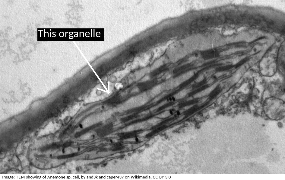
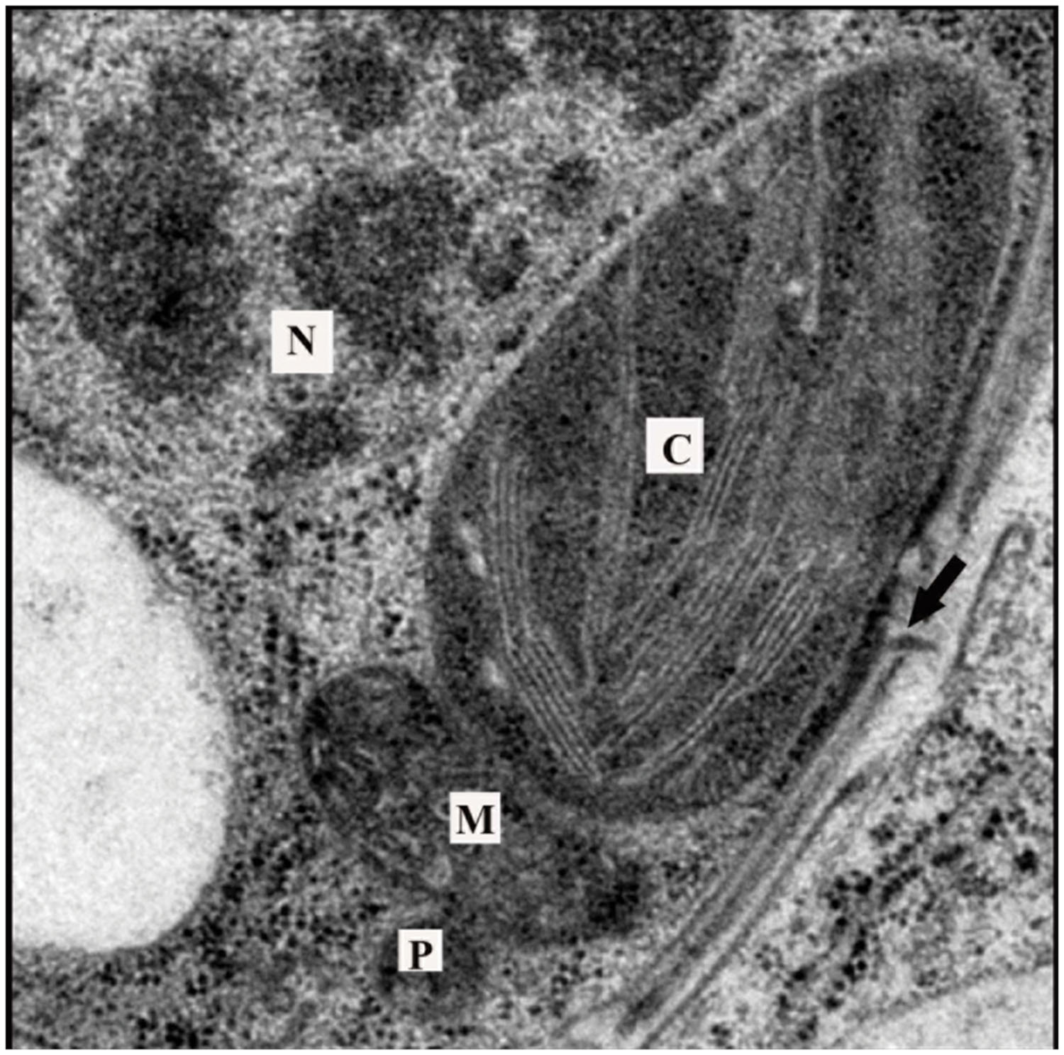
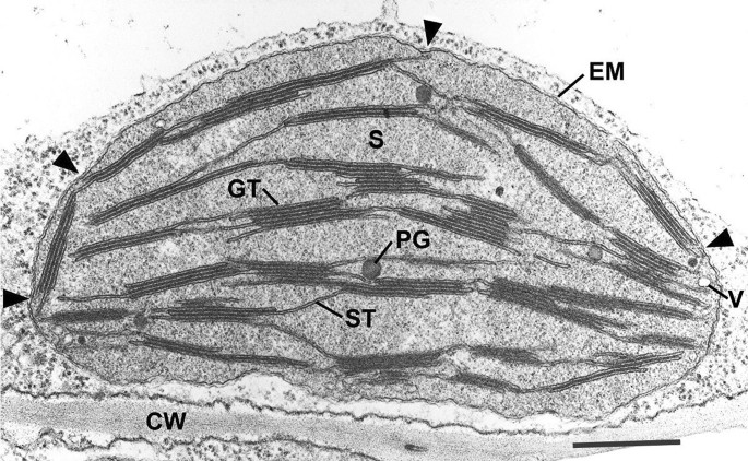
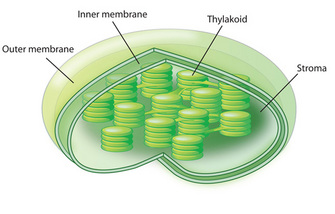
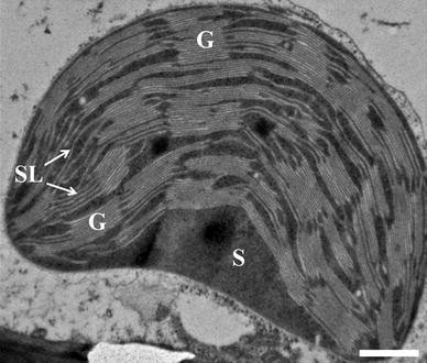
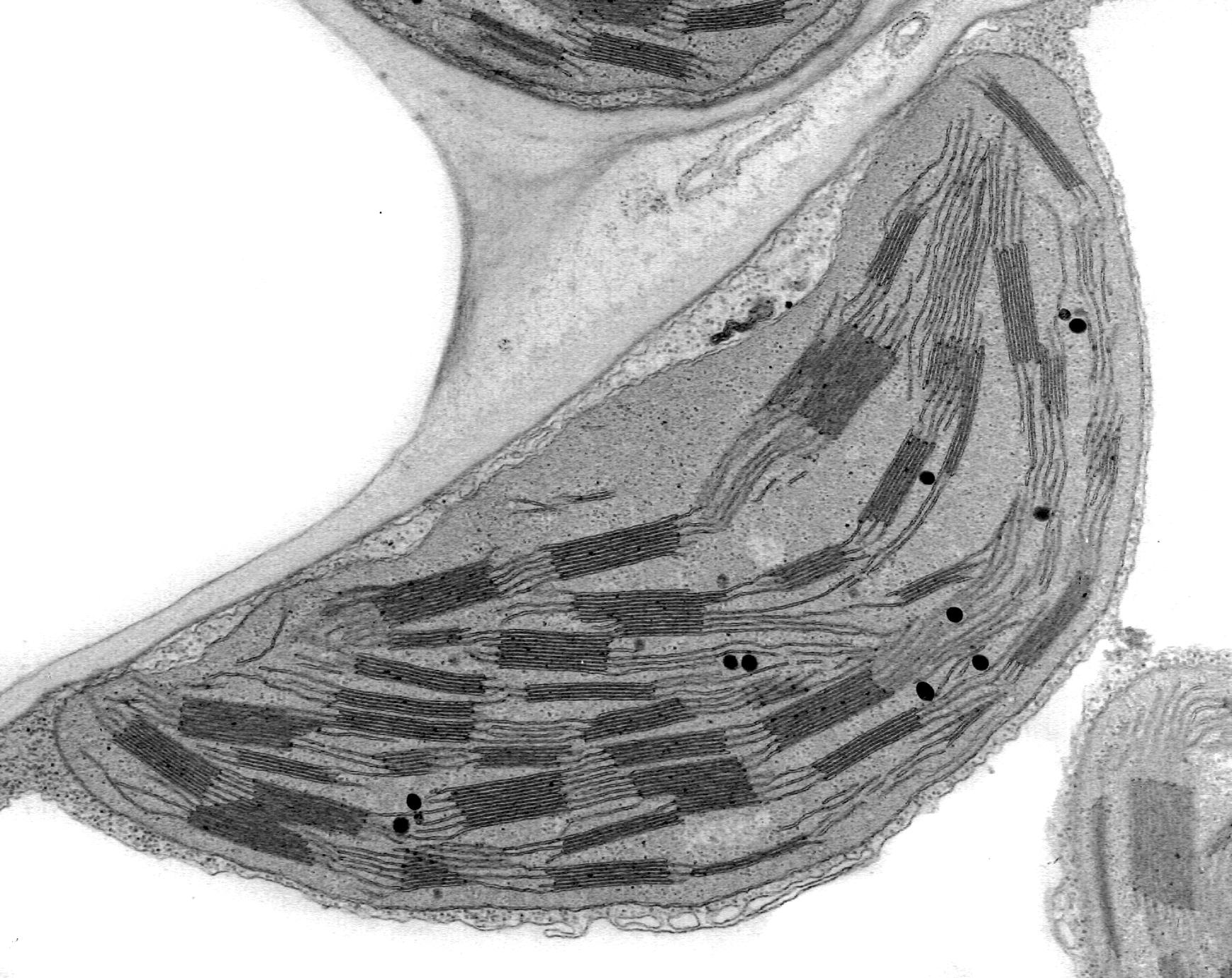
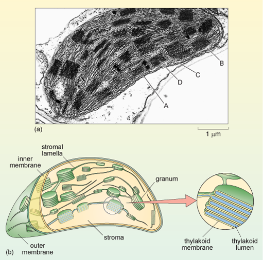
45 label the transmission electron microscope image of a chloroplast below
DP Biology (Cells: 1.1, 1.2, 1.5, 6.3) Quiz - Quizizz The image shows a phagocytic white blood cell as seen with a transmission electron microscope. Which features can be found both within this cell and in a photosynthetic bacterium? DP Biology (Cells: 1.1, 1.2, 1.5, 6.3) DRAFT Looking at the Structure of Cells in the Microscope Images of Surfaces Can Be Obtained by Scanning Electron Microscopy. A scanning electron microscope (SEM) directly produces an image of the three-dimensional structure of the surface of a specimen. The SEM is usually a smaller, simpler, and cheaper device than a transmission electron microscope.
Tumor-resident intracellular microbiota promotes metastatic ... Apr 07, 2022 · High-resolution electron microscopy (EM) analysis showed that the majority of bacteria-like structures were identified in the cytosol rather than the extracellular space (estimated as 97.25% in cytosol, N = 218), and the bacteria-cell ratio was estimated to be 3% (218 bacteria out of 7201 scanned cells) (Figures 2E and S2C). These intracellular ...

Label the transmission electron microscope image of a chloroplast below
PDF Identifying Organelles from an Electron Micrograph - Ms JMO's Biology ... The photograph shown below details chloroplast structure as viewed with a transmission electron microscope Courtesy of Dr. Julian Thorpe - EM & FACS Lab, Biological Sciences University Of Sussex A single Granum Chloroplast envelope visible as two membranes Stroma containing numerous small ribosomes Lamellae connecting different grana Lipid A brief history of how microscopic studies led to the ... - Springer Microscopic studies of chloroplasts can be traced back to the year 1678 when Antonie van Leeuwenhoek reported to the Royal Society in London that he saw green globules in grass leaf cells with his single-lens microscope. Since then, microscopic studies have continued to contribute critical insights into the complex architecture of chloroplast membranes and how their structure relates to ... Lab Manual Exercise # 1a - Palomar College Chloroplast: Membrane-bound organelle and the site of photosynthesis and ATP production in autotrophic plant cells. Like mitochondria, chloroplasts contain their own circular DNA molecules. In fact, chloroplast DNA, including the protein-coding RBCL gene, is often used at the family level to show the relationships between genera and species within plant families.
Label the transmission electron microscope image of a chloroplast below. Electron Microscopy Images - Dartmouth Sep 10, 2021 — We have a library of images recorded using our scanning and transmission electron microscopes. Some are shown below and others elsewhere. ACS Nano | Ahead of Print Articles ASAP (as soon as publishable) are posted online and available to view immediately after technical editing, formatting for publication, and author proofing. photosynthesis.pdf - 228 — Chloroplasts 1. Label the transmission ... 228 — Chloroplasts 1. Label the transmission electron microscope image of a chloroplast below: a) stroma b) stroma lamellae c) outer membrane d) granum e) thylakoid f) inner membrane 2. a) Describe where chlorophyll is found in a chloroplast. In the thylakoid membrane. b) Explain why chlorophyll is found here. Assignment 6, page 2 - NC State University Now, it is time to view a real chloroplast by transmission electron microscopy. Study this transmission electron micrograph of a spinach leaf cell, locate a chloroplast and capture the image for labeling. The micrograph is displayed as if using a "virtual electron microscope", so you will need to magnify the image and move to a region that contains the clearest view of chloroplast internal structures. Perform a screen capture of the chloroplast, then label:
Transmission electron micrographs of armed chlorenchyma cells of O ... Using electron microscopy alone to quantify plasmodesmata is difficult because of the limited surface area coverage per image and hence the need to examine large numbers of sections for robust ... AP Biology Describe the principles, advantages, and limitations of the light microscope, transmission electron microscope, and scanning electron microscope. Electron microscopes - Cell structure - Edexcel - BBC Bitesize the scanning electron microscope (SEM) has a large depth of field. so can be used to examine the surface structure of specimens A human lymphocyte white blood cell as seen with a transmission ... PDF Assignment 6 - NC State University View this transmission electron micrograph of a plant cell, locate a chloroplast and capture the image for labeling. The micrograph is displayed as if using a "virtual electron microscope", so you will need to magnify the image and move to a region that contains the clearest view of chloroplast internal structures. Perform a screen capture of the chloroplast, then label the following structures: a thylakoid
(PDF) The Transmission Electron Microscope 01/01/2009 · Abstract. A typical commercial transmission electron microscope (TEM) costs about 5 for each electron volt (eV) of energy in the beam and, if you add on all available options, it can easily cost ... Chloroplast Electron Microscope - transmission electron … 14/08/2022 · Chloroplast Electron Microscope - 18 images - cell biology chloroplast, molecular make up of cells cells the basic units of life siyavula, chloroplast structure under microscope micropedia, chloroplast under microscope labeled micropedia, Menu ≡ ╳ Home ; Login & Register ; Contact ; Home; Chloroplast Electron Microscope; Chloroplast Electron Microscope. … Chloroplasts - Definition, Structure, Function and Microscopy Microscopy. To view chloroplasts under the microscope, students can use toluidine blue stain to prepare a wet mount. This simply involves the following simple steps: Place a plant sample onto drop of water on a clean glass slide; Using a dropper, add a drop of the stain (toluidine blue) on the sample and allow to stand for about a minute Cell biology - Wikipedia History. Cells were first seen in 17th century Europe with the invention of the compound microscope.In 1665, Robert Hooke termed the building block of all living organisms as "cells" (published in Micrographia) after looking at a piece of cork and observing a cell-like structure, however, the cells were dead and gave no indication to the actual overall components of a cell.
Transmission Electron Microscope (With Diagram) - Biology Discussion Transmission Electron Microscope (With Diagram) In this article we will discuss about the design of transmission electron microscope, explained with the help of a diagram. In TEM a finely focused beam of electrons from an electron gun is passed through a specially prepared ultra thin section of the specimen. The beam is focused on a small area ...
Electron Microscope Principle, Uses, Types and Images (Labeled … 02/02/2022 · Parts of a Transmission Electron Microscope. There are four major components in a transmission electron microscope. They are. Electron Gun; Electromagnetic Lens; Specimen Holder; Image viewer and recording mechanism; Electron Gun: This is the part that generates the electrons. The electron gun houses a tungsten filament that is heated to produce electrons
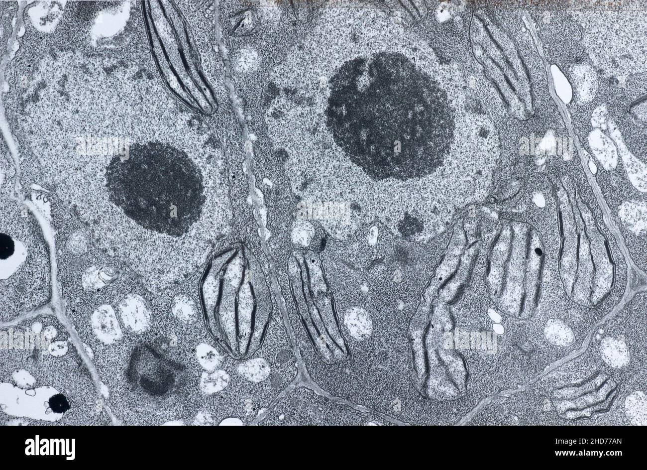
Transmission electron microscopic images of … Transmission electron microscopic images of chloroplasts and mitochondria in 15-day-old leaves from PRORP1 RNAi mutants and wild-type plants. (A, B) Ultrastructure of chloroplasts and mitochondria...
A-level Biology Bridging Course - Week 1 - Amazon S3 The images below are from a scanning electron microscope, transmission electron microscope and a ... (a) Name the parts of the chloroplast labelled A and B.
1. Cell Structure.docx - Q1. The diagram shows part of an... 2. One mark from incorrect answer involving any two hydrogens and an oxygen from carbons 1 and 4; Do not award marks if all atoms concerned are on same carbon atom or are on carbon atoms other than 1 and 4 or where the answer does not have two hydrogen and one oxygen 2 (c) (i) 1. Holds chains / cellulose molecules together / forms cross links between chains / cellulose molecules / forms ...
Use of corpora in translation studies 1137 Projects 1137 incoming 1137 knowledgeable 1137 meanings 1137 σ 1136 demonstrations 1136 escaped 1136 notification 1136 FAIR 1136 Hmm 1136 CrossRef 1135 arrange 1135 LP 1135 forty 1135 suburban 1135 GW 1135 herein 1135 intriguing 1134 Move 1134 Reynolds 1134 positioned 1134 didnt 1134 int 1133 Chamber 1133 termination 1133 overlapping 1132 newborn 1132 Publishers 1132 jazz 1132 Touch 1132 ...
Transmission Electron Microscope (TEM)- Definition, … 19/05/2022 · This is the part of the Transmission Electron Microscope responsible for producing electron beams. Electrons are produced by a …
Course: s4: Biology , Topic: UNIT 3: MICROSCOPY - REB The optical microscope, often referred to as light microscope is a type of microscope which uses visible light and a system of lenses to magnify images of small samples. The different parts of light microscope are described below: -- Base: supports and stabilizes the microscope on the table or any other working place.
Top 14 Types of Spectroscopic Techniques – Explained ... ADVERTISEMENTS: Some of the important types of Spectroscopic Techniques are as follows: Type # 1. Gamma Spectroscopy: Gamma spectroscopy is a radionuclide measurement method. While a Geiger counter determines only the count rate, a gamma spectrometer will determine the energy and the count rate of gamma-rays emitted by radioactive substances. Gamma spectroscopy is an extremely […]
30 Label The Transmission Electron Microscope Image Of A ... Jul 27, 2017 — 30 Label The Transmission Electron Microscope Image Of A Chloroplast Below · Light microscope that provides lightly colored high contrast 3 dim ...
Transmission Electron Microscopy (TEM) for Chloroplast? A Multi-Step Transmission Electron Microscopy Sample Preparation Technique for Indented Ceramics having Extensive Sub-Surface Cracking
Labeling the Cell Flashcards | Quizlet Label the structures of the plasma membrane and cytoskeleton. Label the membranous organelles. ... Label the transmission electron micrograph of the mitochondrion. Label the transmission electron micrograph of the nucleus. membrane bound organelles. golgi apparatus, mitochondrion, lysosome, peroxisome, rough endoplasmic reticulum ...
Solved Part 1 (1 point) In the electron micrograph, identify - Chegg Part 1 (1 point) In the electron micrograph, identify the bacterial cell features by dragging the labels to their targets. Cell wall D A Plasma membrane Cytoplasm Outer membrane B с 1 um Q Courtesy of E. Kellenberger What is a drawback to using light microscopy? Choose one: o O A. It cannot be used to view structures smaller than a bacterium. B.
Ultrastructure of cells quiz 1.2 - Subscription websites for IB ... The electron microscope image below shows an organelle found in both animal and plant cells. ... Chloroplasts are distinctive because they have stacks of membranes inside, called grana, which hold the chlorophyll that absorbs light. ... The production and transmission of a nerve impulse.
I ntroduction to Photosynthesis - Pcmac Based on the information above, label the transmission electron micrograph (TEM) of a chloroplast below: 2. What does chlorophyll do?
Electron Micrographs of Cell Organelles | Zoology - Biology Discussion This is an electron-micrograph of plastid or chloroplast, which is an integral component of all green plant leaves and is characterized by following features (Fig. 15 & 16): (1) They may be spheroidal, ovoid, stellate or collar shaped and differ in size and number in different cells.
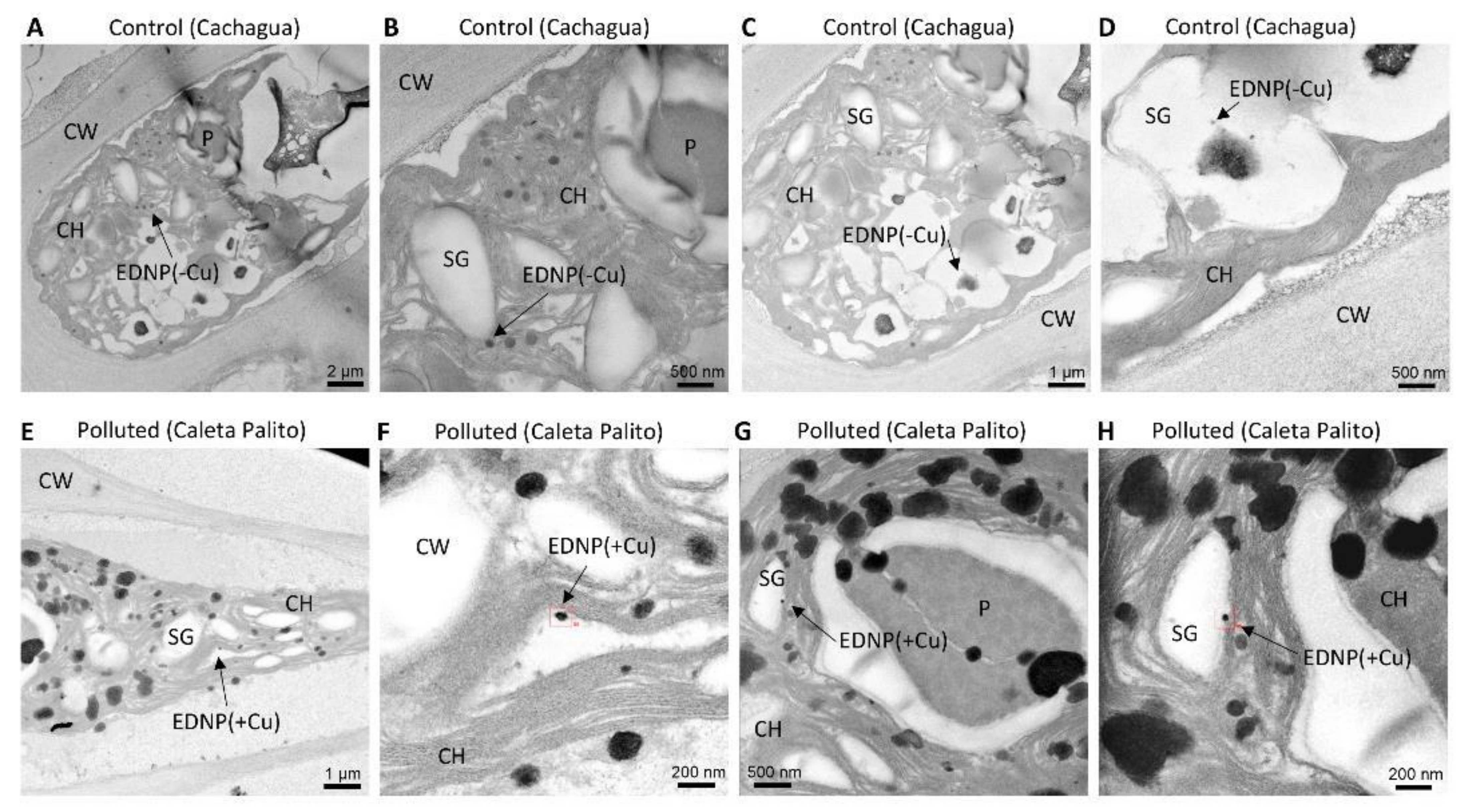
Chlorophyll catabolism precedes changes in chloroplast structure and ... Transmission electron microscopy (TEM) of chemically fixed leaf samples from the four developmental stages revealed the ultrastructural changes within chloroplasts as they senesce. Most noticeable are the decrease in the size of chloroplasts, disappearance of starch bodies, increase in number and size of plastoglobules (PGs), and disintegration of the thylakoid membranes (Figure 3 a-l).
Tumor-resident intracellular microbiota promotes metastatic ... Apr 14, 2022 · High-resolution electron microscopy (EM) analysis showed that the majority of bacteria-like structures were identified in the cytosol rather than the extracellular space (estimated as 97.25% in cytosol, N = 218), and the bacteria-cell ratio was estimated to be 3% (218 bacteria out of 7201 scanned cells) (Figures 2E and S2C).
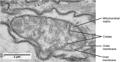
Cells and magnification Flashcards | Quizlet 6) a- Figure 2 shows a photos of part of a mitochondrion from a mouse liver cell taken using a transmission electron microscope at x62800 magnification Produce a scientific drawing of the mitochondria in figure 2 Label the following part of the mitochondrion on your drawing -Matrix-Crista (4 marks)
The Transmission Electron Microscope | CCBER - UC … Transmission electron microscopes (TEM) are microscopes that use a particle beam of electrons to visualize specimens and generate a highly-magnified image. TEMs can magnify objects up to 2 million times. In order to get a better idea of just how small that is, think of how small a cell is. It is no wonder TEMs have become so valuable within the ...
Field emission scanning electron microscopy of microtubule arrays in ... Summary Specimen preparation protocols that allow field emission scanning electron microscope imaging of microtubules in plant cells were developed, involving simultaneous permeabilization with saponin and stabilization of microtubules with taxol.
Electron Microscope- Definition, Principle, Types, Uses, Labeled Diagram There are two types of electron microscopes, with different operating styles: 1. Transmission Electron Microscope (TEM) The transmission electron microscope is used to view thin specimens through which electrons can pass generating a projection image. The TEM is analogous in many ways to the conventional (compound) light microscope.
What are the labels of the transmission electronic microscope ... What are the labels of the transmission electronic microscope image of a chloroplast - 24307282 rsc96fd8wc rsc96fd8wc 07/28/2021 Biology High School answered What are the labels of the transmission electronic microscope image of a chloroplast 1 See answer Advertisement Advertisement rsc96fd8wc is waiting for your help. Add your answer and earn ...
Cambridge International AS & A Level Biology Coursebook ... 1.6 Plant and animal cells as seen with an electron microscope w ie id br ev The fine (detailed) structure of a cell as revealed by the electron microscope is called ultrastructure and is shown in Figures 1.18–1.21.-R s ve rs Question op y Copy and complete Table 1.2, which compares light microscopes with electron microscopes.
Chloroplast - Wikipedia Scanning transmission electron microscope imaging of a chloroplast (Top) 10-nm-thick STEM tomographic slice of a lettuce chloroplast. Grana stacks are interconnected by unstacked stromal thylakoids, called "stroma lamellae". Round inclusions associated with the thylakoids are plastoglobules. Scalebar = 200 nm. See.
Assignment 6, page 1 Now, it is time to view a chloroplast by transmission electron microscopy. View this transmission electron micrograph of a plant cell , locate a chloroplast and capture the image for labeling. The micrograph is displayed as if using a "virtual electron microscope", so you will need to magnify the image and move to a region that contains the clearest view of chloroplast internal structures.
Chloroplast Electron Microscope - transmission electron micrograph of ... Chloroplast Electron Microscope - 18 images - chloroplast structure under microscope micropedia, cell biology chloroplast, light and electron microscope images ccber, labled diagram of a chloroplast micrograph photo by akucic biology,
Lab Manual Exercise # 1a - Palomar College Chloroplast: Membrane-bound organelle and the site of photosynthesis and ATP production in autotrophic plant cells. Like mitochondria, chloroplasts contain their own circular DNA molecules. In fact, chloroplast DNA, including the protein-coding RBCL gene, is often used at the family level to show the relationships between genera and species within plant families.
A brief history of how microscopic studies led to the ... - Springer Microscopic studies of chloroplasts can be traced back to the year 1678 when Antonie van Leeuwenhoek reported to the Royal Society in London that he saw green globules in grass leaf cells with his single-lens microscope. Since then, microscopic studies have continued to contribute critical insights into the complex architecture of chloroplast membranes and how their structure relates to ...
PDF Identifying Organelles from an Electron Micrograph - Ms JMO's Biology ... The photograph shown below details chloroplast structure as viewed with a transmission electron microscope Courtesy of Dr. Julian Thorpe - EM & FACS Lab, Biological Sciences University Of Sussex A single Granum Chloroplast envelope visible as two membranes Stroma containing numerous small ribosomes Lamellae connecting different grana Lipid





![PDF] Occurrence and forms of Kranz anatomy in photosynthetic ...](https://d3i71xaburhd42.cloudfront.net/58a9714e4b3303ed24ec52f0b25fc553fd1cec27/7-Figure5-1.png)




![PDF] Transgenic plants : methods and protocols | Semantic Scholar](https://d3i71xaburhd42.cloudfront.net/94df879997abffa87e171bd2fee15fbd30945268/131-Figure4-1.png)






Post a Comment for "45 label the transmission electron microscope image of a chloroplast below"