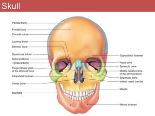
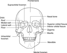
40 figure 14.1 label the anterior bones and features of the skull
Solved Parietal one)... Frontal Coronel Suture, Labore bo ... - Chegg Frontal Coronel Suture, Labore bo- - Supra ortal foc MEN - Nasal Tampopal port osphenoid FO FIGURE 14.1 of that bone) 1 Label the anterior bones and features of the skull of the line lacks the word bone, label the particular feature KPR Donel bore) bonel Ethmoid bone (bone) bone) bone) (bone) (bone) FIGURE 14.2 Label the lateral bones and Material properties of the human cranial vault and zygoma Occipital bone sites O2-O5 have unique features, including some of the thickest cortical bone in the skull. The distinct material properties of the occipital bone may relate to its functional role as an attachment area for the nuchal musculature, which functions in head posturing and movement.
Craniosynostosis in the Middle Pleistocene human Cranium 14 from the ... Relevant features in Cranium 14. (A) Posterior view, showing the parallelogram profile and the ipsilateral occipito-mastoid bulge, both diagnostic features of the left lambdoid suture premature fusion.Note the deviation of the sagittal plane with respect to the sagittal suture plane, showing that the Inion, the occipital crest and other medial structures of the nuchal plane are positioned 10 ...
Figure 14.1 label the anterior bones and features of the skull
BW AP Skull fig 14.1 lab - Figure 14.1 Label the anterior bones and ... ANATOMY AN ANATOMY AN 101 BW AP Skull fig 14.1 lab - Figure 14.1 Label the anterior bones and features of the skull. (If the line lacks the word bone, label the particular BW AP Skull fig 14.1 lab - Figure 14.1 Label the anterior... School University of Kentucky Course Title ANATOMY AN 101 Type Lab Report Uploaded By christine97 Pages 1 Skeletal elements of the penguin eye and their functional and ... In birds, it is reinforced by both bone and cartilage. The bony element consists of a ring of scleral ossicles as well as, unique among vertebrates to some birds, the os opticus (os nervi optici, Gemminger's ossicle). The scleral ossicles are a ring of interlocking bones found at the anterior margin of the sclera near the corneo‐scleral junction. 6.3 Bone Structure - Anatomy and Physiology 2e | OpenStax Bones of the pelvis, skull, spine, and legs are the most commonly affected. When occurring in the skull, Paget's disease can cause headaches and hearing loss. Figure 6.14 Paget's Disease Normal leg bones are relatively straight, but those affected by Paget's disease are porous and curved. What causes the osteoclasts to become overactive?
Figure 14.1 label the anterior bones and features of the skull. A&P 1 Lab Figure 14.1 Bones & Features of the skull (anterior view ... Start studying A&P 1 Lab Figure 14.1 Bones & Features of the skull (anterior view). Learn vocabulary, terms, and more with flashcards, games, and other study tools. 19.3 Joints and Skeletal Movement - Concepts of Biology - 1st Canadian ... Sutures are found only in the skull and possess short fibers of connective tissue that hold the skull bones tightly in place (Figure 19.23). ... Protraction is the anterior movement of a bone in the horizontal plane. Retractionoccurs as a joint moves back into position after protraction. Protraction and retraction can be seen in the movement of ... PDF 'l-f,------ - That Science Life Figure 14.1 Label the anterior bones and features of the skull. (If the line lacks the word bone,label the particular feature of that bone .) (bone) .,' -z(bone) ^ Lacrimal bone Ethmoid bone (bone) Sphenoid bone (bone) (bone) (bone) (bone) 14 14 16 (bone) Fi.gure 14.2 Label the lateral bones and features of the skull. (bone) .'' /hnna\ \vv' Lab 14: Figure 14.3 Inferior Bones of the Skull - Quizlet Lab 14: Figure 14.3 Inferior Bones of the Skull STUDY Learn Write Test PLAY Match Created by THagge TEACHER Terms in this set (15) Zygomatic Arch ... Vomer Bone ... Temporal Bone ... Maxilla ... Palatine Process of Maxilla ... Zygomatic Bone ... Palatine Bone ... Sphenoid Bone ... Styloid Process ... Exteral Acoustic Meatus ... Occipital Process
Vertebral Column and Thoracic Cage Lab - Studyres Complete Parts A, B, C, and D. 1 Figure 14.1 Label the bones and features of the lateral view (left) and the posterior view (right) of the vertebral column. Figure 14.2 Label the superior views of the atlas and axis. 2 Figure 14.3 Label the features of the cervical, thoracic, and lumbar vertebrae. 3 Figure 14.4 Label the coccyx and the features ... Systematics of Eosemionotus The left hyomandibula is isolated and displaced posterodorsal to the skull in MCSN 8006 (Figure 6.2, Figure 9). The bone has a relatively narrow and slightly ventrally expanded shaft, with the hyomandibular foramen close to the anterior border, and a posteriorly expanded dorsal portion, but there is no distinct opercular process. 19.1 Types of Skeletal Systems - Concepts of Biology - 1st Canadian Edition The human pectoral girdle consists of the clavicle (or collarbone) in the anterior, and the scapula (or shoulder blades) in the posterior (Figure 19.11). Figure 19.11. (a) The pectoral girdle in primates consists of the clavicles and scapulae. (b) The posterior view reveals the spine of the scapula to which muscle attaches. Visit wwwmhhecommartinseries1 for pre lab questions - Course Hero (if the line lacks the word bone, label the particular feature of that bone.) 1 2occipital bone (1) lambdoid suture external occipital protuberance foramen magnum occipital condyle temporal bone (2) squamous suture external acoustic meatus mandibular fossa mastoid process styloid process carotid canal jugular foramen internal acoustic meatus …
16.5 Musculoskeletal System - Concepts of Biology | OpenStax The joints between the bones in the skull and between the teeth and the bone of their sockets are examples of fibrous joints (Figure 16.16a). Cartilaginous joints are joints in which the bones are connected by cartilage (Figure 16.16b). An example is found at the joints between vertebrae, the so-called "disks" of the backbone. Solved bone) (bone From the provided list, enter in each - Chegg Transcribed image text: bone) (bone From the provided list, enter in each blank below the term that properly identifies each labeled part of the skull. As usual, your answer must be entered exactly as shown below in order to get credit. Use each term once and only once. coronal suture Lacrimal bone - frontal Ethmoid bone inferior nasal concha mandible bone) maxilla Sphenoid bone middle nasal ... 7.1 Divisions of the Skeletal System - OpenStax The axial skeleton of the adult consists of 80 bones, including the skull, the vertebral column, and the thoracic cage. The skull is formed by 22 bones. Also associated with the head are an additional seven bones, including the hyoid bone and the ear ossicles (three small bones found in each middle ear). Lab 14: Figure 14.1 Anterior Bones of the Skull Diagram | Quizlet Lab 14: Figure 14.1 Anterior Bones of the Skull STUDY Learn Write Test PLAY Match + − Created by THagge TEACHER Terms in this set (16) Parietal ... Frontal ... Coronal Suture ... Supraorbital Foramen ... Nasal Bone ... Temporal Bone ... Perpendicular Plate of the Ethmoid Bone ... Infraorbital Foramen ... Vomer Bone ... Mandible ... Mental Foramen
Skull and C-Spine Lab Images from Lab Manual - Figure 14.1 Label the ... ANATOMY AN ANATOMY AN 101 Skull and C-Spine Lab Images from Lab Manual - Figure 14.1 Label the anterior bones and features of the skull. (If the line lacks the word bone, label Skull and C-Spine Lab Images from Lab Manual - Figure 14.1... School University of Kentucky Course Title ANATOMY AN 101 Type Lab Report Uploaded By christine97 Pages 4
CH 14 HW.pdf - 4/21/2021 CH 14 HW CH 14 HW Due: 11:59pm on... - Course Hero 4/21/2021 CH 14 HW 1/12 CH 14 HW Due: 11:59pm on Monday, April 12, 2021 To understand how points are awarded, read the Grading Policy for this assignment. Art Labeling Activity: Figure 14.1 Part A Label the parts of the upper respiratory system (1 of 2). Drag the labels onto the diagram to identify the parts of the upper respiratory system (1 of 2). ANSWER: Correct Art Labeling Activity ...
PDF Laboratory Manual for Human Anatomy & Physiology Fetal Pig Version, 4e ... Revised and added leader lines and labels New figures Revised and expanded components 13 Pre-Lab Figure 13.2 (bone features) Added questions Added labels 14 Pre-Lab Procedure (skull) Figure 14.1, 14.2, 14.4, 14.5, 14.11 (skull) Figure 14.3 (mandible) Figure 14.11 (lateral view of skull) Assessments: Part A and Part D Added questions
7.2 The Skull - Anatomy and Physiology 2e | OpenStax The anterior skull consists of the facial bones and provides the bony support for the eyes and structures of the face. This view of the skull is dominated by the openings of the orbits and the nasal cavity. Also seen are the upper and lower jaws, with their respective teeth ( Figure 7.4 ).
Intervertebral disc - Physiopedia The intervertebral disc (IVD) is important in the normal functioning of the spine. It is a cushion of fibrocartilage and the principal joint between two vertebrae in the spinal column. There are 23 discs in the human spine: 6 in the cervical region (neck), 12 in the thoracic region (middle back), and 5 in the lumbar region (lower back).
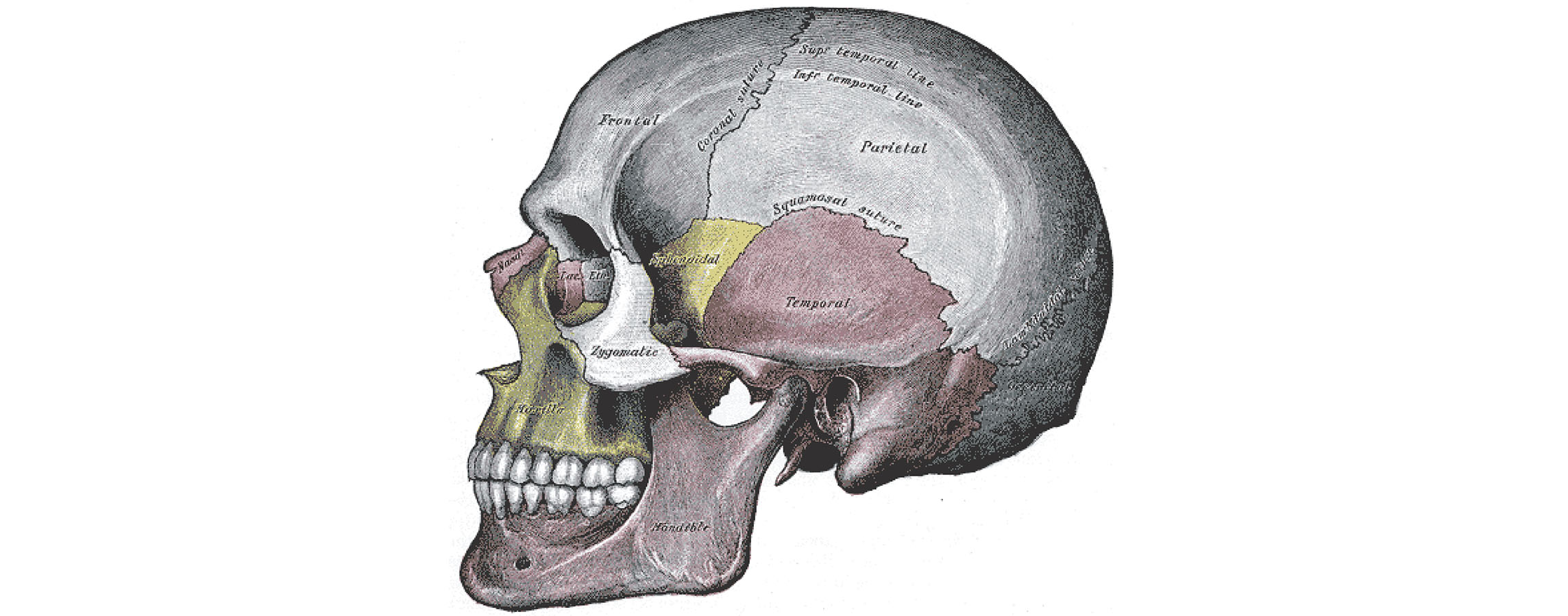
Lab 14: Figure 14.11 Lateral View of the Skull Diagram | Quizlet Lab 14: Figure 14.11 Lateral View of the Skull STUDY Learn Write Test PLAY Match + − Created by THagge TEACHER Terms in this set (14) Parietal ... Squamosal Suture ... Temporal Bone ... Lambdoidal Suture ... Occipital Bone ... External Acoustic Meatus ... Coronal Suture ... Frontal Bone ... Zygomatic Process ... Zygomatic Bone ... Maxilla ...
PDF Laboratory Manual for Human Anatomy & Physiology Cat Version, 4e 13 Pre-Lab Figure 13.2 (bone features) Added questions Added labels 14 Pre-Lab Procedure (skull) Figure 14.1, 14.2, 14.4, 14.5, 14.11 (skull) Figure 14.3 (mandible) Figure 14.11 (lateral view of skull) Assessments: Part A and Part D Added questions Revised and added components Added labels New figure Added term to label Added questions
Lab exercise 12.1, 14.1 ,14.2 anthropology - SlideShare Measure and calculate indices for a Neanderthal and an anatomically modern human skull. Fill out the table below as you work. a. Calculate the cranial index. Cranial breadth * 100 = Cranial Index Cranial length b. Use the sliding calipers to measure the comparative length of the anterior and posterior tooth rows.
Musculoskeletal System - Bone Development Timeline Introduction. The adult human skeleton has about 206 different bones, each develop with their own specific bone timeline. Many prenatal bones fuse postnatal developing neonate and child (about 275). The two main forms of ossification occur in different bones, intramembranous (eg skull) and endochondral (eg vertebra) ossification.
1.6 Anatomical Terminology - Anatomy and Physiology 2e - OpenStax The anterior (ventral) cavity has two main subdivisions: the thoracic cavity and the abdominopelvic cavity (see Figure 1.15). The thoracic cavity is the more superior subdivision of the anterior cavity, and it is enclosed by the rib cage. The thoracic cavity contains the lungs and the heart, which is located in the mediastinum.
6.3 Bone Structure - Anatomy and Physiology 2e | OpenStax Bones of the pelvis, skull, spine, and legs are the most commonly affected. When occurring in the skull, Paget's disease can cause headaches and hearing loss. Figure 6.14 Paget's Disease Normal leg bones are relatively straight, but those affected by Paget's disease are porous and curved. What causes the osteoclasts to become overactive?
Skeletal elements of the penguin eye and their functional and ... In birds, it is reinforced by both bone and cartilage. The bony element consists of a ring of scleral ossicles as well as, unique among vertebrates to some birds, the os opticus (os nervi optici, Gemminger's ossicle). The scleral ossicles are a ring of interlocking bones found at the anterior margin of the sclera near the corneo‐scleral junction.
BW AP Skull fig 14.1 lab - Figure 14.1 Label the anterior bones and ... ANATOMY AN ANATOMY AN 101 BW AP Skull fig 14.1 lab - Figure 14.1 Label the anterior bones and features of the skull. (If the line lacks the word bone, label the particular BW AP Skull fig 14.1 lab - Figure 14.1 Label the anterior... School University of Kentucky Course Title ANATOMY AN 101 Type Lab Report Uploaded By christine97 Pages 1






Post a Comment for "40 figure 14.1 label the anterior bones and features of the skull"