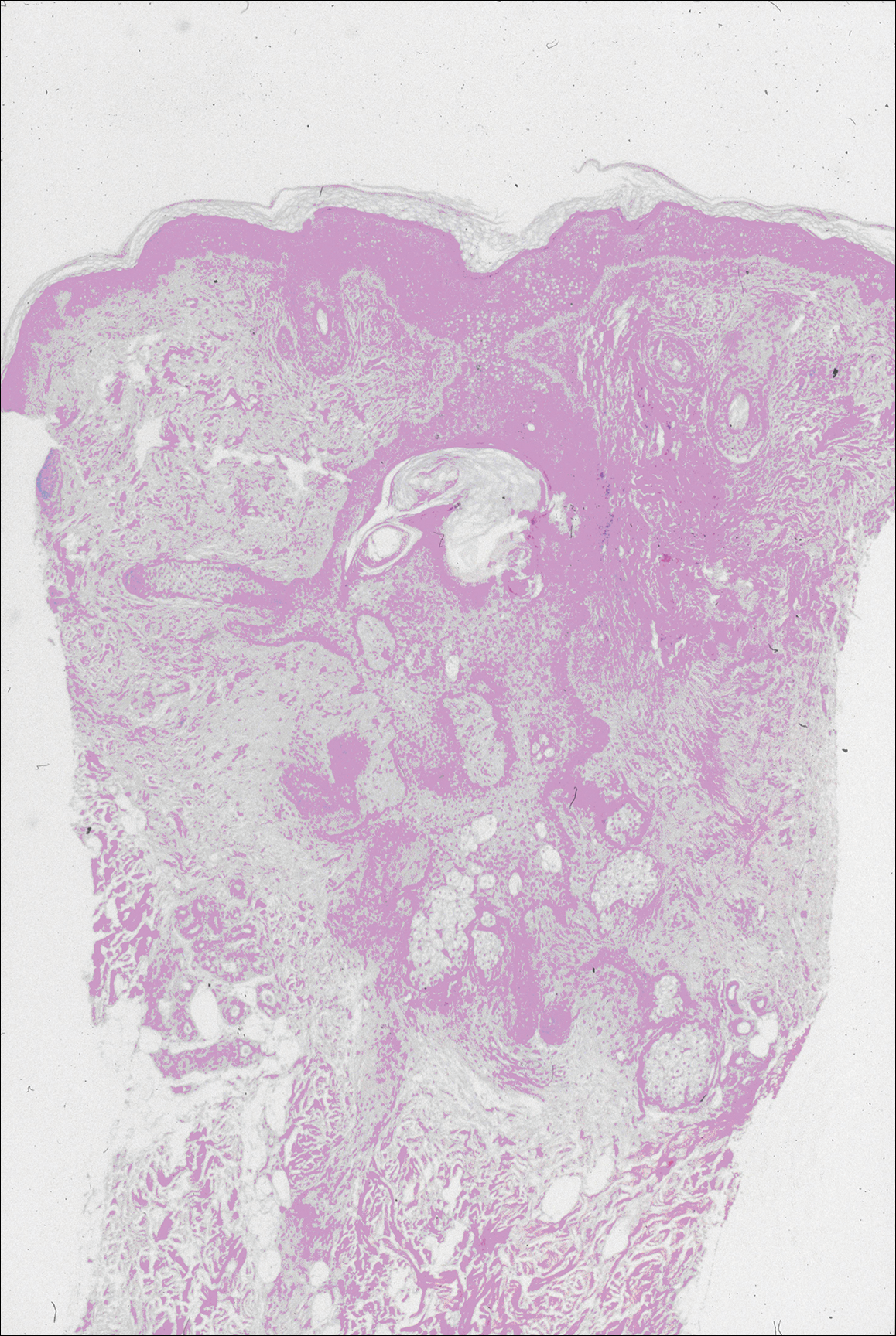
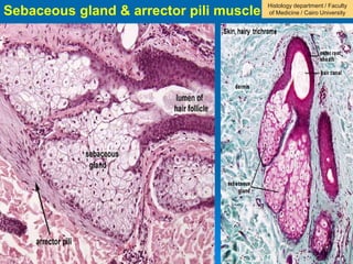
38 label the photomicrograph of the sebaceous gland
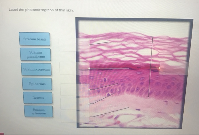
Label The Photomicrograph Of Thin Skin Quizlet / 290 Anatomy Ideas ... In the photomicrograph shown above, which layer do new cells arise? C) contains more sweat glands than thin skin. Label the photomicrograph of thin skin. In the photomicrograph of a portion of thick skin shown below, . 28) in the diagram of skin shown below, which labeled structure generates fingerprints? Label the photomicrograph of thin skin ... Label The Photomicrograph Of Thin Skin And Its Accessory Structures ... The skin and its accessory structures make up the integumentary system, which provides the. Epidermis sebaceous gland hair follicle duct of sebaceous gland . The nail bed is a . Hair is made of dead keratinized cells, and gets its color from . Label the photomicrograph of the skin and its accessory structures.
Highly Persistent Label-Retaining Cells in the Hair Follicles of Mice ... Slowly cycling, [3 H]thymidine label-retaining cells (LRC) have been observed after short intervals of 4-10 wk following [3 H]thymidine labeling of baby mice or of adult mice after repeated treatment with 12-O-tetradecanoylphorbol-13-acetate, to reside in the middle third of the hair follicle immediately beneath the sebaceous gland near the ...
Label the photomicrograph of the sebaceous gland
Anatomy and Physiology Homework Chapter 6 Flashcards | Quizlet Consider sebaceous and ceruminous glands. Then click and drag each label into the appropriate category based on whether it pertains to sebaceous glands, ceruminous glands, or both.-Usually opens up into a hair follicle-Secretes sebum-Coats guard hairs to improve their performance-Coats the scalp hair with oil-Blockage and infection can cause pimples HelenakruwAllison Label the Photomicrograph of the Sebaceous Gland. These patients often complain of. ... Saya pun pada mulanya ingatkan sama saja tapi selepas mengu… Total Pageviews Powered by Blogger Labels 2 2017 5 a Agar Air an Apa Bagi Beli Berbuka Best Buat Car Cara Cek Combustion Contoh Daerah Dan Deals Described Detox Di English Floors Gland Hari ... Label The Photomicrograph Of Thin Skin And Its Accessory Structures ... The nail bed is a . Name the 4 layers of thin skin in both the cartoon and the photomicrograph. The skin and its accessory structures make up the integumentary system,. Identify the layers, cells and structures as indicated. Sebaceous gland duct of sebaceous gland epidermis hair follicle . Apocrine and merocrine (eccrine) glands are .
Label the photomicrograph of the sebaceous gland. (Solved) - Label the photomicrograph of thin skin. Label the ... Thin Skin Histology HM 44 Label The Photomicrograph Of Thin Skin. Points 01:04:56 Stratum Granulosum EBook Stratum Spinosum Dermis Epidermis Stratum Corneum Stratum Basale (Solved) - Label the photomicrograph of thin skin. Dermis Duct of ... The Integumentary System A. Anatomy of the skin. Identify and label the photograph of the skin model: epidermis, dermis, dermal papillae, hypodermis, hair follicle, hair bulb, sebaceous gland, sudoriferous gland, duct of sudoriferous gland, pore of... Chapter 6 - Labeling Parts of Skin - sweat gland blood... sweat gland blood vessels sweat sweat gland pore muscle layer Labeling Parts of the Skin Identify the layers of skin. dermis stratum basale stratum spinosum stratum lucidum stratum corneum stratum granulosum basement membrane Identify the parts of this photomicrograph of skin: sebaceous gland hair shaft hair follicle dermis epidermis Question : Label the photomicrograph of the sebaceous gland. - Chegg Experts are tested by Chegg as specialists in their subject area. We review their content and use your feedback to keep the quality high. 100% (28 ratings) Transcribed image text: Label the photomicrograph of the sebaceous gland. Previous question Next question.
Label The Photomicrograph Of Thin Skin. - Skin Model 1 - YouTube Hair sebaceous gland dermis hair follicle epidermis duct of sebaceous. Some labeled features may be referred to once, more than once, or not at all. ... Hair sebaceous gland dermis hair follicle epidermis duct of sebaceous. Label the photomicrograph of thin skin. This is a photomicrograph of thin skin. Learn more about thin skin treatment at ... Photomicrograph Of Thick Skin Labeled : Integument Sciencedirect Learn vocabulary, terms, and more with flashcards, games, and other study tools. 1 answer to label the photomicrograph of thin skin. Learn about dermatitis and how you can treat it Take several photomicrographs of thin skin at this magnification. ... Thick skin has a thinner dermis than thin skin, and does not contain hairs, sebaceous glands ... Label the Photomicrograph of the Sebaceous Gland. Solved Label The Photomicrograph Of The Sebaceous Gland Chegg Com Organize the following layers of the epidermis from superficial to deep.. The skin consists of two layers. Boys can also blame testosterone from gonadal puberty pubarche. Use Enter Space to view and traverse through the list of languages. -Photomicrograph of sebaceous glands at the periphery of the glandular ... Download scientific diagram | -Photomicrograph of sebaceous glands at the periphery of the glandular welt from the specimen in Fig. 4. A, x95; B, holocrine-secreting cells of sebaceous glands ...
Sebaceous Gland Label The Photomicrograph Of Thin Skin - This ... This micrograph shows layers of skin in a cross section from oerpub.github.io Sebaceous gland arrector pili muscle merocrine sweat gland hair. 1 answer to label the photomicrograph of thin skin. Using the slide thin skin with hairs, and the photomicrographs of cutaneous glands (figure 7.7) as . Label the photomicrograph of thin skin. Photomicrograph Of Thick Skin Labeled : Jaypeedigital Ebook Reader A few layers of cells that are . Label the photomicrograph of thick skin. Stratum corneum stratum basale stratum granulosum stratum lucidum epidermis dermis stratum . Learn vocabulary, terms, and more with flashcards, games, and other study tools. Thick skin has a thinner dermis than thin skin, and does not contain hairs, sebaceous glands, . Figure 7.4 Photomicrograph of the skin and accessory structures a projection of connective tissue into the hair follicle and contains blood vessels that provide nutrients to the dividing cells of the matrix. Sebaceous Gland. Oil glands that surround hair follicles; secrete oils that lubricates skin, hair, and into the neck of the hair follicle. Hair Follicle. Label The Photomicrograph Of Thin Skin Quizlet - Photomicrograph ... Epidermis, hair, duct, sebaceous, hair, dermis. Label the photomicrograph of thin skin. Label the photomicrograph of thin skin. Start studying photomicrographs of thin skin. D) stratum corneum has fewer layers in. In the photomicrograph of a portion of thick skin shown below, . Label the structures of the skin and subcutaneous tissue.
STRUKTUR DAN FUNGSI JARINGAN SERTA ORGAN
[Solved] C ezto.mheducation.com/hm.tpx 23. Label the photomicrograph of ... Label the photomicrograph of thin skin. Hair Sebaceous gland Dermis Hair Follicle Epidermis Duct of sebaceous. Study Resources. Main Menu; by School; by Literature Title; ... Hair Sebaceous gland Dermis Hair Follicle Epidermis Duct of sebaceous gland KS Name the structure. Type here to search T... Biology Science Anatomy. Comments (0) ...

Human scalp, light micrograph - Stock Image - C054/3494 ...
Sebaceous Gland Label The Photomicrograph Of Thin Skin ... - Blogger Sebaceous Gland Label The Photomicrograph Of Thin Skin / Accessory Structures Of The Skin Anatomy And Physiology. Friday, February 18, 2022. Human, rat, pig, cow and … chex %parser=2.13 %floated=19991204 %generated=dr/all %bound=true This is the spellchex dictionary for online spell checking. ...

A&P 1 Exercise_7 Activity 1 & 2 & RYK and UYK.docx - LAB ...
Question : Label the photomicrograph of thin skin. Dermis Duct of ... Transcribed image text: Label the photomicrograph of thin skin. Dermis Duct of sebaceous gland Hair Follicle Sebaceous gland Hair Epidermis
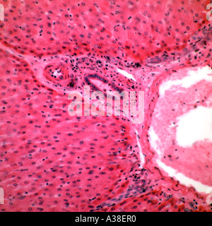
Human liver section brightfield photomicrograph Stock Photo ...
Label The Photomicrograph Of Thin Skin Quizlet : 35 Label The ... Label the structures of the skin and subcutaneous tissue. Epidermis, hair, duct, sebaceous, hair, dermis. Label the photomicrograph of thin skin. In the diagram of skin shown below, which structure illustrates that thin. What is this region of skin called (the general name.not any of the specific layers found within this region)?. 35 Label The ...
Bioengineering in Wound Healing : Anatomy of the Human Skin ...
Sebaceous Gland - an overview | ScienceDirect Topics Sebaceous glands are more numerous along the dorsum in dogs and sparse along the ventrum—an important factor to consider when sampling for sebaceous adenitis. ... Photomicrograph of Zymbal's gland tumor showing the typical mixed squamous (arrow) and sebaceous differentiation. The arrowhead depicts a nest of plump neoplastic sebaceous ...

Visor Redalyc - Warthin-like mucoepidermoid carcinoma of the ...
Label The Photomicrograph Of Thin Skin And Its Accessory Structures ... The nail bed is a . Name the 4 layers of thin skin in both the cartoon and the photomicrograph. The skin and its accessory structures make up the integumentary system,. Identify the layers, cells and structures as indicated. Sebaceous gland duct of sebaceous gland epidermis hair follicle . Apocrine and merocrine (eccrine) glands are .
Co-occurrence of Steatocystoma Multiplex, Eruptive Vellus ...
HelenakruwAllison Label the Photomicrograph of the Sebaceous Gland. These patients often complain of. ... Saya pun pada mulanya ingatkan sama saja tapi selepas mengu… Total Pageviews Powered by Blogger Labels 2 2017 5 a Agar Air an Apa Bagi Beli Berbuka Best Buat Car Cara Cek Combustion Contoh Daerah Dan Deals Described Detox Di English Floors Gland Hari ...

Photomicrographs of skin. (a) Dog 1. Sebaceous glands are ...
Anatomy and Physiology Homework Chapter 6 Flashcards | Quizlet Consider sebaceous and ceruminous glands. Then click and drag each label into the appropriate category based on whether it pertains to sebaceous glands, ceruminous glands, or both.-Usually opens up into a hair follicle-Secretes sebum-Coats guard hairs to improve their performance-Coats the scalp hair with oil-Blockage and infection can cause pimples
Solved Label these structures located in axillary skin. Hair ...
Integumentary System Overview

Hamza Khan (captainhamzakhan0) - Profile | Pinterest
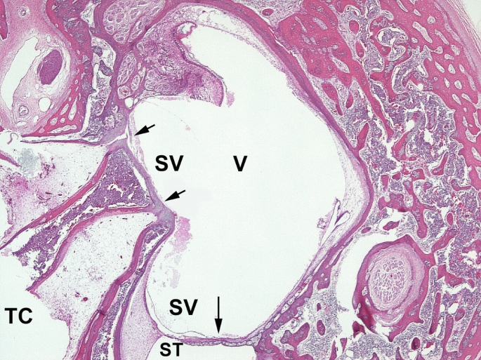
Ear | SpringerLink

Chapter 6 Study Set Flashcards | Quizlet

Secretory Tissue Photos - Free & Royalty-Free Stock Photos ...

Photomicrograph of hair follicle surrounded by sebaceous ...

The photomicrograph shows a sebaceous carcinoma demonstrating ...

Sebaceous hyperplasia of the vulva: a clinicopathological ...

Cherenfant_iLab 4.docx - BIO251/Section 7 Week 4 Lab ...

sebaceous gland tissue - Keyword Search - Science Photo Library
Co-occurrence of Steatocystoma Multiplex, Eruptive Vellus ...

Figure 7.4: Photomicrograph of the skin and accessory ...

Lap Practical #1 EC Flashcards | Quizlet

7,937 Tissue Anatomy Photos and Premium High Res Pictures ...

Photomicrograph of hair follicle surrounded by sebaceous ...
Histology Submandibular Gland Type Salivary Gland Stock Photo ...
Skin and the Integumentary System
Mucocele and Ranula Workup: Imaging Studies, Procedures ...
Solved Label the photomicrograph of the skin and its | Chegg.com

The photomicrograph of the rat skin stained with Heidenhain's ...
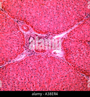
Human liver section brightfield photomicrograph Stock Photo ...
Astragalus membranaceus and Punica granatum alleviate ...

JaypeeDigital | eBook Reader
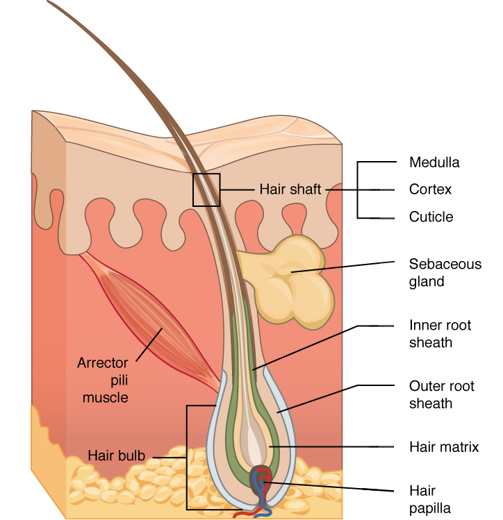
Hair | Biology for Majors II
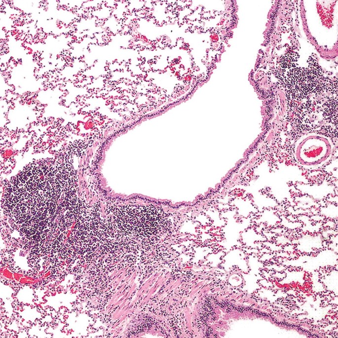
Malignant Hematopoietic Disorders of the Lung | SpringerLink
integumentary system
ANATOMY OF THE HUMAN EYE: November 2005
Sebaceous gland hi-res stock photography and images - Alamy

a): A photomicrograph of the section of thin skin tissue from ...
Post a Comment for "38 label the photomicrograph of the sebaceous gland"