44 eye diagram not labeled
Eye Anatomy: Parts of the Eye and How We See Behind the anterior chamber is the eye's iris (the colored part of the eye) and the dark hole in the middle called the pupil. Muscles in the iris dilate (widen) or constrict (narrow) the pupil to control the amount of light reaching the back of the eye. Directly behind the pupil sits the lens. The lens focuses light toward the back of the eye. Diagram of the Eye - Home - Lions Eye Institute To understand the eye and its functions, it's important to understand how the eye works, see below diagrams for both the external eye and the internal eye. The External Eye Instructions Click the parts of the eye to see a description for each. Hover the diagram to zoom. The Internal Eye Instructions
Labelling the eye - Science Learning Hub Use your mouse or finger to hover over a box to highlight the part to be named. Drag and drop the text labels onto the boxes next to the eye diagram If you want to redo an answer, click on the box and the answer will go back to the top so you can move it to another box. If you want to check your answers, use the 'Reset incorrect' button.
Eye diagram not labeled
PDF Anatomy of an Eye Diagram - Tektronix Figure 6. An eye diagram triggered from a clock recovered from the data signal using a narrow loop bandwidth clock recovery scheme. Figure 5. An incomplete eye diagram formed by triggering on data. Figure 8. An eye diagram triggered such that the delay between jittered clock and jittered data destructively interferes. Figure 7. Anatomy of the Eye Diagrams for Coloring/Labeling, with Reference and ... Some of these eye diagrams could be printed on an overhead, or included in a higher level test. There are blank versions, so you can customize it for your specific needs. Complete Package Contents: - One colored and labeled reference chart, showing the cross-sectional anatomy of the eye, (Not for black and white photocopying). Anatomy of an Eye Diagram: How to Construct & Trigger Mask testing is an abbreviated eye diagram test for the quick testing of transmitters in manufacturing. Rather than measuring all parametric aspects of the eye, mask testing defines key areas in the eye that are deemed to be "keep-out" areas - if any of the signal is detected to be in this region, then the device fails.
Eye diagram not labeled. Optic vesicle - Wikipedia The eyes begin to develop as a pair of diverticula (pouches) from the lateral aspects of the forebrain.These diverticula make their appearance before the closure of the anterior end of the neural tube; after the closure of the tube around the 4th week of development, they are known as the optic vesicles.Previous studies of optic vesicles suggest that the surrounding extraocular … Eye Anatomy: 16 Parts of the Eye & Their Functions Apr 05, 2022 · The conjunctiva is the membrane covering the sclera (white portion of your eye). The conjunctiva also covers the interior of your eyelids. Conjunctivitis, often known as pink eye, occurs when this thin membrane becomes inflamed or swollen. Other eye disorders that affect the conjunctiva include: The Eye Diagram: What is it and why is it used? The eye diagram is used primarily to look at digital signals for the purpose of recognizing the effects of distortion and finding its source. To demonstrate using a Tektronix MDO3104 oscilloscope, we connect the AFG output on the back panel to an analog input channel on the front panel and press AFG so a sine wave displays. Then we press Acquire. Eye Diagram With Labels and detailed description - BYJUS A brief description of the eye along with a well-labelled diagram is given below for reference. Well-Labelled Diagram of Eye The anterior chamber of the eye is the space between the cornea and the iris and is filled with a lubricating fluid, aqueous humour. The vascular layer of the eye, known as the choroid contains the connective tissue.
Parts of a microscope with functions and labeled diagram 19/04/2022 · Q. Differentiate between a condenser and an Abbe condenser. Ans. Condensers are lenses that are used to collect and focus light from the illuminator into the specimen. They are found under the stage next to the diaphragm of the microscope. They play a major role in ensuring clear sharp images are produced with a high magnification of 400X and above. Human Eye - Definition, Structure, Function, Parts, Diagram Structure of Human Eye. A human eye is roughly 2.3 cm in diameter and is almost a spherical ball filled with some fluid. It consists of the following parts: Sclera: It is the outer covering, a protective tough white layer called the sclera (white part of the eye). Cornea: The front transparent part of the sclera is called the cornea. Microscope, Microscope Parts, Labeled Diagram, and Functions 19/01/2022 · Revolving Nosepiece or Turret: Turret is the part of the microscope that holds two or multiple objective lenses and helps to rotate objective lenses and also helps to easily change power. Objective Lenses: Three are 3 or 4 objective lenses on a microscope. The objective lenses almost always consist of 4x, 10x, 40x and 100x powers. The most common eyepiece lens is … Block Diagram | Complete Guide with Examples - Edraw 08/12/2021 · Not only this, but a well-prepared block diagram also gives you an overview of the expected outcomes of the process you are planning to implement. Therefore, here you will learn what block diagrams are and what their advantages are, what points must be kept in mind while preparing a block diagram , and how to prepare one in the easiest possible manner with the …
Eye Diagram: Label Quiz - PurposeGames.com A Rorschach Test 13p Image Quiz. Match the fractions, percentage and decimals 8p Matching Game. Spot 5 differences 5p Shape Quiz. Passed the eye test? 10p Image Quiz. Continue to count 15p Multiple-Choice. •. •. •. Eye Diagram Quiz - ProProfs Try this amazing Eye Diagram Quiz quiz which has been attempted 4326 times by avid quiz takers. Also explore over 77 similar quizzes in this category. Take Quizzes. Animal; Nutrition; ... Quiz: Label The Parts Of The Eye. People say that the eyes are the windows to a person's soul. In the class today, we covered parts of the eye, and what ... Human eye - Wikipedia The human eye is a sensory organ, part of the sensory nervous system, that reacts to visible light and allows us to use visual information for various purposes including seeing things, keeping our balance, and maintaining circadian rhythm.. The eye can be considered as a living optical device.It is approximately spherical in shape, with its outer layers, such as the outermost, white … The Human Eye (Eyeball) Diagram, Parts and Pictures The eyeball is a round gelatinous organ that contains the actual optical apparatus. It is approximately 25 mm in diameter and sits snugly in the orbit where six muscles control its movement. The eyeball has three layers, each of which has several important structures that are essential for the sense of vision. Wall of the Eyeball
Lab 3 Sensory Physiology
Compound Microscope- Definition, Labeled Diagram, Principle, … 03/04/2022 · Therefore, a 10x eyepiece used with a 40X objective lens will produce a magnification of 400X. The naked eye can now view the specimen at magnification 400 times greater and so microscopic details are revealed. Alternatively, the magnification of the compound microscope is given by: m = D/ f o * L/f e

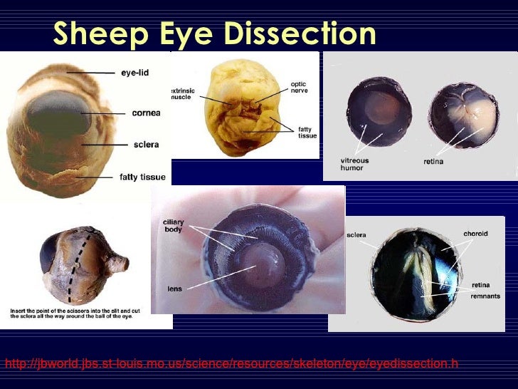
Sheep Eye Dissection - YouTube
Labelled Diagram of Human Eye, Explanation and Function - VEDANTU The human eye is a part of the sensory nervous system. Labeled Diagram of Human Eye The eyes of all mammals consist of a non-image-forming photosensitive ganglion within the retina which receives light, adjusts the dimensions of the pupil, regulates the availability of melatonin hormones, and also entertains the body clock.

Labeled Eye Diagram
Labeled Eye Diagram | Science Trends What you want to interpret as a major part of the human eye is somewhat up to the individual, but in general there are seven parts of the human eye: the cornea, the pupil, the iris, the lens, the vitreous humor, the retina, and the sclera. Let's take a closer look at each of these components individually. The Cornea
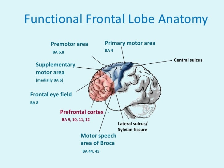
Frontal lobe
Iris (eye) - Simple English Wikipedia, the free encyclopedia The iris (plural: irides or irises) is a thin, circular structure in the eye. It controls the diameter and size of the pupils. Eye colour is the colour of the iris. In humans, the iris may look green, blue, brown, hazel (a combination of light brown, green and gold), grey, violet, or even pink. In response to the amount of light exiting the eye ...

Human A&P: Anatomy of the Eye - YouTube
Eye Diagram Unlabelled - Wiring Diagram Pictures Jul 12, 2018 · Select the correct label for each part of the eye. The image is taken from above the left eye. Click on the Score button to see how you did. Incorrect answers will. 61 high-quality Unlabeled Eye Diagram for free! human eye diagram human eye diagram Watch on Download and use them in your website, document or presentation.

Gentry, Teresa M / Anatomy Diagrams
CUT-AND-ASSEMBLE PAPER EYE MODEL THE HUMAN EYE 1) OPTIC NERVE: takes electrical signals to the brain. Notice that the retina’s blood supply comes in through the center of the optic nerve. 2) FOVEA: focal point, the center of your vision 3) MACULA: the area around the fovea 4) RETINA: the back of the inside of the eyeball (This is where the light-sensitive rods and cones are located.)

Labeling the Eyeball Quiz
Eye Anatomy Diagram - EnchantedLearning.com Aqueous humor - the clear, watery fluid inside the eye. It provides nutrients to the eye. Astigmatism - a condition in which the lens is warped, causing images not to focus properly on the retina. Binocular vision - the coordinated use of two eyes which gives the ability to see the world in three dimensions - 3D. Cones - cells the in the retina that sense color.
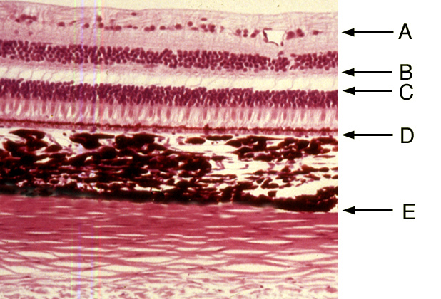
Eye | histology
Eye diagram basics: Reading and applying eye diagrams - EDN Figure 3 (a) Improper termination causes an eye diagram to look stressed. (b) Proper termination relaxes the eye. As can be seen in Figure 4, an eye diagram can reveal important information. It can indicate the best point for sampling, divulge the SNR (signal-to-noise ratio) at the sampling point, and indicate the amount of jitter and distortion.
Post a Comment for "44 eye diagram not labeled"